Non-compression oncological myelopathies
- Understanding Non-Compressive Oncological Myelopathies
- Types of Non-Compressive Oncological Myelopathies
- Diagnosis of Non-Compressive Oncological Myelopathies
- Treatment and Management Principles
- Differential Diagnosis of Non-Compressive Myelopathy in Cancer Patients
- Prognosis and Complications
- When to Consult a Specialist
- References
Understanding Non-Compressive Oncological Myelopathies
Myelopathies (disorders of the spinal cord) in patients with malignant tumors most commonly arise from compressive lesions, such as vertebral metastases leading to epidural spinal cord compression. However, a subset of cancer patients develops spinal cord dysfunction due to non-compressive mechanisms. These non-compressive oncological myelopathies can be challenging to diagnose and differentiate from compressive causes or other neurological conditions.
Overview and Diagnostic Challenges
When imaging studies such as MRI or CT of the spinal cord (including myelography) do not reveal evidence of external spinal cord compression or a block in cerebrospinal fluid (CSF) flow, it becomes difficult to distinguish between various non-compressive causes. These include intramedullary metastases (metastases within the spinal cord parenchyma), paraneoplastic myelopathies (spinal cord damage as a remote effect of cancer, not due to direct tumor invasion or metastasis), and radiation myelopathy (damage from prior radiation therapy).
If a patient with known cancer develops progressive myelopathy and imaging (myelography, CT, or MRI) shows no signs of external spinal cord compression, intramedullary metastasis is a primary consideration. Paraneoplastic myelopathy occurs much less frequently. A patient's complaint of back pain may be the first symptom of an intramedullary metastasis, but this is not a mandatory or universal finding. With intramedullary metastases, patients may develop progressive spastic paraparesis (weakness and increased tone in the legs) or paresthesias (abnormal sensations like tingling or numbness).
Dissociated sensory loss (e.g., loss of pain and temperature sensation with preserved light touch and proprioception) or complete preservation of sensation in the sacral segments (sacral sparing) are characteristic features of intrinsic (intramedullary) spinal cord compression or lesions. However, these classic patterns are rarely observed in their pure form in patients with intramedullary metastases. More commonly, asymmetric paraparesis and partial or patchy loss of sensation are encountered.
When a patient is diagnosed using myelography, CT, or MRI, spinal cord edema may be visible even without signs of external compression. It's important to note that in up to half of spinal cord examinations in patients using CT and myelography, a normal picture might be seen, especially in early or subtle non-compressive myelopathies. Therefore, MRI of the spinal cord is considered the most informative diagnostic method for differentiating an intramedullary metastatic focus from a primary intramedullary tumor or other non-compressive conditions.
Types of Non-Compressive Oncological Myelopathies
Intramedullary Metastases
Tumor metastases within the spinal cord parenchyma (intramedullary metastases) are relatively rare compared to epidural metastases causing cord compression. They usually originate from primary cancers such as bronchogenic carcinoma (lung cancer), less frequently from breast cancer, and other solid tumors. Melanoma metastases, while rarely causing external spinal cord compression, are more commonly found as intramedullary tumors. An intramedullary metastasis typically appears as a single, often eccentrically located, nodule within the spinal cord, formed as a result of hematogenous (bloodstream) spread of cancer cells. Radiation therapy for melanoma metastasis in the spinal cord may offer some palliative effect or local control, but the overall prognosis is often poor.
Carcinomatous Meningitis (Leptomeningeal Carcinomatosis)
Carcinomatous meningitis, also known as leptomeningeal carcinomatosis or neoplastic meningitis, is a common form of central nervous system (CNS) involvement in cancer. It occurs when cancer cells spread to the leptomeninges (the arachnoid and pia mater, the inner membranes covering the brain and spinal cord) and the cerebrospinal fluid (CSF). Carcinomatous meningitis may not directly cause myelopathy unless the cancerous infiltration spreads from the adjacent nerve roots under the arachnoid membrane to involve the spinal cord surface or superficial parenchyma. Alternatively, it can lead to the formation of tumor nodules on the surface of the cord or within the nerve root sleeves, causing secondary compression or direct infiltration of the spinal cord.
An incomplete cauda equina syndrome, often not accompanied by a significant pain syndrome, can be caused by carcinomatous infiltration of the nerve roots forming the cauda equina. Patients with carcinomatous meningitis frequently complain of headaches, cranial nerve palsies, and radicular symptoms. Serial analyses of CSF in such patients often reveal malignant cells (positive cytology), increased protein content, and a decrease in glucose concentration (hypoglycorrhachia).
In suspected carcinomatous meningitis, a lumbar puncture (spinal tap) is performed to obtain cerebrospinal fluid (CSF). Analysis of the CSF for malignant cells, protein levels, and glucose concentration is crucial for diagnosis.
Paraneoplastic Necrotizing Myelopathy
Paraneoplastic necrotizing myelopathy is a rare neurological syndrome that occurs as a remote, non-metastatic effect of cancer, typically associated with solid tumors (e.g., lung cancer, ovarian cancer, lymphoma). It involves progressive necrosis (tissue death) of the spinal cord, often accompanied by mild inflammation. The exact pathogenesis is not fully understood but is thought to be immune-mediated, possibly involving autoantibodies targeting neural antigens.
Clinical presentation usually involves a subacute progressive spastic paraparesis that develops over several days or weeks, often characterized by asymmetry. This is typically accompanied by paresthesias in the distal extremities, which can ascend to form a distinct sensory level. Dysfunction of the pelvic organs (bladder and bowel dysfunction) often occurs later in the course. Paraneoplastic necrotizing myelopathy commonly affects several adjacent spinal cord segments, leading to a longitudinally extensive transverse myelitis-like picture. Myelographic findings and routine CSF analysis are often normal or may show only a slight increase in protein levels. Specific onconeural antibodies may sometimes be detected in serum or CSF.
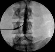
Myelography, a diagnostic imaging technique where contrast agent is injected into the subarachnoid space (historically referred to as subdural in some contexts, but correctly subarachnoid) via lumbar puncture, can visualize the outline of the spinal cord and detect compressive lesions or blocks in CSF flow. Modern practice often uses CT myelography or MRI.
Radiation Myelopathy
Radiation therapy administered for oncological diseases can cause a delayed, progressive myelopathy if the spinal cord is included within the radiation field. This is often a serious differential diagnostic challenge for neurosurgeons or neurologists, particularly when evaluating new neurological symptoms in a cancer patient who has previously received radiation to areas near the spine (e.g., for mediastinal lymph nodes, head and neck cancers, or vertebral metastases).
Radiation myelopathy is typically caused by radiation-induced damage to small blood vessels (vasculopathy, hyalinization, and occlusion) within the spinal cord, leading to chronic ischemia and demyelination, or direct radiation damage to oligodendrocytes and neural tissue. It usually manifests months to years after radiation therapy (latent period can range from 6 months to several years, sometimes longer).
The clinical presentation is often a progressive, non-acute myelopathy with symptoms such as spastic weakness, sensory disturbances (paresthesias, sensory level), and sometimes Lhermitte's sign (electric shock-like sensation down the spine or limbs on neck flexion). Differentiating radiation myelopathy from paraneoplastic myelopathy or intramedullary metastasis can be very difficult based on clinical symptoms alone, especially if the primary cancer is still present or has recurred. A clear history of previous radiation therapy with the spinal cord within the treatment port is crucial information. MRI findings in radiation myelopathy can include spinal cord swelling, T2 hyperintensity, and sometimes contrast enhancement in the irradiated segment, but these can also mimic tumor or inflammation.
Diagnosis of Non-Compressive Oncological Myelopathies
The diagnostic approach aims to identify the specific cause of myelopathy in a cancer patient when overt spinal cord compression is absent:
- Neurological Examination: To define the pattern and severity of neurological deficits (motor, sensory, autonomic) and localize the level of spinal cord involvement.
- Magnetic Resonance Imaging (MRI) of the Spine with Gadolinium Contrast: This is the most critical imaging study.
- For intramedullary metastases, MRI typically shows one or more enhancing lesions within the spinal cord parenchyma, often with surrounding edema.
- For carcinomatous meningitis, MRI may show diffuse or nodular leptomeningeal enhancement along the surface of the spinal cord, cauda equina, or nerve roots.
- For paraneoplastic necrotizing myelopathy, MRI may show longitudinally extensive T2 hyperintense signal changes within the cord, sometimes with patchy enhancement or cord swelling, but often without a discrete mass.
- For radiation myelopathy, MRI may show T2 hyperintensity and swelling in the irradiated segment of the cord, sometimes with contrast enhancement. Findings can be non-specific.
- Cerebrospinal Fluid (CSF) Analysis (via Lumbar Puncture):
- Essential for diagnosing carcinomatous meningitis (detection of malignant cells via cytology, elevated protein, low glucose).
- In paraneoplastic myelopathy, CSF may show mild pleocytosis or elevated protein, and specific onconeural antibodies may be present.
- In intramedullary metastases or radiation myelopathy, CSF is often non-specific but can show elevated protein.
- Search for Primary Cancer or Metastases Elsewhere: If the primary cancer is unknown or if new neurological symptoms develop, staging studies (e.g., CT chest/abdomen/pelvis, PET scan) are important to identify the primary tumor or other metastatic sites.
- Electrophysiological Studies (EMG/NCS, Evoked Potentials): Can help characterize the extent of neurological damage and rule out peripheral neuropathy.
- Biopsy (Rarely): Biopsy of a suspected intramedullary lesion or leptomeningeal involvement is rarely performed due to high risk, but may be considered in exceptional cases if diagnosis remains elusive and treatment implications are significant.
Treatment and Management Principles
Treatment depends on the specific type of non-compressive oncological myelopathy:
- Intramedullary Metastases: Treatment options include radiation therapy (often whole-cord or involved-field), corticosteroids (to reduce edema), and sometimes surgical resection for solitary, accessible lesions, particularly if causing significant neurological deficit or if diagnosis is uncertain. Systemic chemotherapy targeted at the primary cancer may also have some effect. Prognosis is generally poor.
- Carcinomatous Meningitis: Treatment involves intrathecal chemotherapy (administered directly into the CSF), systemic chemotherapy that penetrates the CNS, and/or radiation therapy to symptomatic sites. Corticosteroids are used to manage symptoms. Prognosis is generally poor.
- Paraneoplastic Necrotizing Myelopathy: Treatment is challenging. Options include immunosuppressive therapy (corticosteroids, IVIG, plasma exchange, rituximab), and treatment of the underlying cancer. Neurological recovery is often limited.
- Radiation Myelopathy: There is no definitive cure. High-dose corticosteroids are often tried, but their efficacy is limited. Supportive care and management of symptoms (e.g., spasticity, pain) are crucial. Prevention by limiting spinal cord radiation dose during cancer treatment is key.
- Supportive Care: For all types, comprehensive supportive care is essential, including pain management, rehabilitation (physical and occupational therapy), management of bladder/bowel dysfunction, prevention of complications like pressure sores and DVT, and psychosocial support.
Differential Diagnosis of Non-Compressive Myelopathy in Cancer Patients
It is vital to differentiate these oncological myelopathies from other conditions that can cause spinal cord dysfunction in cancer patients, especially those that are treatable or have a different prognosis:
| Condition | Key Differentiating Features |
|---|---|
| Intramedullary Metastasis | Known primary cancer, MRI shows enhancing lesion(s) within cord parenchyma. CSF may have elevated protein. |
| Carcinomatous Meningitis | Headache, cranial nerve palsies, radicular symptoms, myelopathy. CSF cytology positive for malignant cells, low glucose, high protein. MRI may show leptomeningeal enhancement. |
| Paraneoplastic Necrotizing Myelopathy | Subacute progressive myelopathy, often longitudinally extensive. MRI may show T2 hyperintensity, swelling. CSF often mild inflammation. Onconeural antibodies may be present. Occurs as remote effect of cancer. |
| Radiation Myelopathy | History of radiation therapy with spinal cord in field. Delayed onset (months to years). Progressive myelopathy. MRI shows changes in irradiated segment. |
| Epidural Spinal Cord Compression (ESCC) | Back pain, progressive neurological deficits. MRI/CT clearly shows extradural mass (e.g., vertebral metastasis) compressing the cord. This is a compressive myelopathy. |
| Transverse Myelitis (non-oncological) | Acute/subacute onset. May be idiopathic, post-infectious, or related to autoimmune disease (e.g., MS, NMOSD). MRI shows intrinsic cord lesion. No evidence of cancer directly causing it. |
| Spinal Cord Infarction | Sudden onset, often with pain. Characteristic vascular territory involvement on MRI (DWI positive acutely). May be related to hypercoagulability in cancer patients or aortic disease. |
| Nutritional/Metabolic Myelopathy (e.g., B12 deficiency) | Subacute onset. Posterior and lateral column signs. Can occur in cancer patients due to malnutrition or malabsorption. Specific deficiencies identified by lab tests. |
Prognosis and Complications
The prognosis for most non-compressive oncological myelopathies is generally poor, reflecting the advanced nature of the underlying cancer or the irreversible damage caused by radiation or paraneoplastic processes.
- Intramedullary Metastases: Median survival is often measured in months, though radiation can provide temporary stabilization or improvement.
- Carcinomatous Meningitis: Prognosis is typically poor, with median survival of a few months even with treatment.
- Paraneoplastic Necrotizing Myelopathy: Often results in severe, permanent neurological disability. Response to immunosuppression is variable.
- Radiation Myelopathy: Usually progressive and irreversible. Management is supportive.
Complications are primarily related to the progressive neurological deficits, including paralysis, sensory loss, intractable pain, bladder/bowel/sexual dysfunction, pressure sores, DVT/PE, and respiratory failure.
When to Consult a Specialist
Any cancer patient who develops new or worsening neurological symptoms suggestive of spinal cord dysfunction requires urgent evaluation by a neurologist and/or neurosurgeon, often in consultation with their oncologist. Key symptoms warranting immediate attention include:
- Progressive weakness or numbness in the limbs.
- Difficulty walking or maintaining balance.
- New onset of bowel or bladder dysfunction.
- New or worsening back pain, especially if associated with neurological symptoms.
- Sensory level (a clear line below which sensation is altered).
Prompt diagnosis and differentiation from compressive myelopathy (which may be more readily treatable with surgical decompression) are critical for guiding appropriate management and providing prognostic information.
References
- Posner JB. Neurologic Complications of Cancer. 2nd ed. Oxford University Press; 2005. (Seminal textbook)
- Schiff D, Kesari S, Wen PY. Cancer Neurology in Clinical Practice: Neurologic Complications of Cancer and its Treatment. 2nd ed. Humana Press; 2008.
- Costigan DA, Winkelman MD. Intramedullary spinal cord metastasis. A clinicopathological study of 13 cases. J Neurosurg. 1985 Aug;63(2):227-33.
- Chamberlain MC. Neoplastic meningitis. Neurologist. 2008 May;14(3):174-84.
- Dropcho EJ. Paraneoplastic neurological syndromes. Handb Clin Neurol. 2012;105:583-602.
- Schultheiss TE, Kun LE, Ang KK, Stephens LC. Radiation response of the central nervous system. Int J Radiat Oncol Biol Phys. 1995 Jul 15;31(5):1093-112.
- Kaley TJ, DeAngelis LM. Therapy of L CNS metastases. Arch Neurol. 2009 Jun;66(6):710-4. (Relevant to carcinomatous meningitis)
- Cole JS. Spinal cord tumors. Curr Treat Options Neurol. 2008 Mar;10(2):109-19. (Context for intramedullary lesions)
See also
- Anatomy of the spine
- Ankylosing spondylitis (Bechterew's disease)
- Back pain by the region of the spine:
- Back pain during pregnancy
- Coccygodynia (tailbone pain)
- Compression fracture of the spine
- Dislocation and subluxation of the vertebrae
- Herniated and bulging intervertebral disc
- Lumbago (low back pain) and sciatica
- Osteoarthritis of the sacroiliac joint
- Osteocondritis of the spine
- Osteoporosis of the spine
- Guidelines for Caregiving for Individuals with Paraplegia and Tetraplegia
- Sacrodinia (pain in the sacrum)
- Sacroiliitis (inflammation of the sacroiliac joint)
- Scheuermann-Mau disease (juvenile osteochondrosis)
- Scoliosis, poor posture
- Spinal bacterial (purulent) epiduritis
- Spinal cord diseases:
- Spinal spondylosis
- Spinal stenosis
- Spine abnormalities
- Spondylitis (osteomyelitic, tuberculous)
- Spondyloarthrosis (facet joint osteoarthritis)
- Spondylolisthesis (displacement and instability of the spine)
- Symptom of pain in the neck, head, and arm
- Pain in the thoracic spine, intercostal neuralgia
- Vertebral hemangiomas (spinal angiomas)
- Whiplash neck injury, cervico-cranial syndrome


