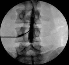
Discography, myelography
Myelography: Imaging the Spinal Canal
Principle and Purpose
Myelography is an invasive diagnostic imaging procedure that involves the introduction of a contrast medium into the subarachnoid space (the space containing cerebrospinal fluid or CSF) surrounding the spinal cord and nerve roots within the dural sac. The primary purpose of myelography is to visualize the CSF-filled spaces and detect abnormalities that may be impinging upon or deforming the spinal cord or nerve roots. This technique allows for the identification of pathological formations, spinal cord deformities, mechanical obstacles to CSF flow, and blockages of cerebrospinal fluid pathways.
It is particularly useful for evaluating conditions like spinal stenosis, herniated intervertebral discs, spinal tumors, cysts, arteriovenous malformations, and nerve root impingement, especially when MRI is contraindicated or provides insufficient detail.
Traditional Contrast Myelography Procedure
The technical execution of traditional myelography, while requiring precision, is generally not overly complex for experienced practitioners. The procedure typically involves:
- Lumbar Puncture: A lumbar puncture (spinal tap) is performed under sterile conditions, usually at the L3–L4 or L4–L5 vertebral level in the lumbar spine. The patient is often positioned prone or on their side.
- Contrast Agent Injection: After confirming correct needle placement in the subarachnoid space (indicated by free flow of CSF), a small amount of CSF may be removed for analysis, and then a contrast agent is injected.
- Historically, gas (such as oxygen or nitrous oxide, creating a negative contrast) or oil-based contrast media were used.
- Modern practice primarily utilizes water-soluble, non-ionic iodinated contrast agents (e.g., Amipaque (metrizamide - historical), Dimer-X (meglumine iocarmate - historical), Omnipaque (iohexol), Isovue (iopamidol)). These agents mix with the CSF and outline the spinal structures.
- Imaging: Following contrast injection, X-ray images (fluoroscopy and plain radiographs) are taken as the patient is tilted on the table to allow the contrast to flow to different levels of the spinal canal (cervical, thoracic, lumbar).
During a traditional myelogram, a contrast agent is injected into the subarachnoid space (often incorrectly referred to as subdural in lay terms, but correctly the subarachnoid space) via a lumbar puncture. This allows visualization of the spinal cord and nerve roots on subsequent X-ray imaging.
Modern Advancements: CT Myelography and MR Myelography
Today, traditional plain X-ray myelography is often followed by a Computed Tomography (CT) scan (CT Myelography) while the contrast is still in the subarachnoid space. CT myelography provides much greater detail and cross-sectional images, significantly enhancing diagnostic accuracy compared to plain film myelography alone.
Furthermore, a non-invasive method, Magnetic Resonance Myelography (MR Myelography), is now widely available. For MRI scans of the spinal cord, special heavily T2-weighted sequences can be used to create myelographic-like images that highlight the CSF spaces without the need for a lumbar puncture or contrast injection. This non-injectable method allows assessment of the degree of filling of the subarachnoid space and its patency for cerebrospinal fluid flow. The resulting images are often three-dimensional, facilitating visualization and diagnosis of the level and nature of lesions for neurosurgeons and radiologists.
A three-dimensional MR myelogram demonstrating the lumbar spinal canal, thecal sac, and exiting nerve roots, achieved non-invasively using specialized MRI sequences.
Indications for Myelography
Myelography (conventional, CT, or MR) is typically considered when:
- MRI is contraindicated (e.g., patient has an incompatible pacemaker or certain metallic implants).
- MRI results are inconclusive or do not correlate with clinical findings.
- Detailed visualization of nerve root sleeves or subtle CSF leaks is required.
- Evaluation of spinal canal stenosis, herniated discs, spinal tumors, cysts, or arteriovenous malformations.
- Assessment of post-traumatic or post-surgical changes in the spinal canal.
- Planning for spinal surgery or interventions.
Contraindications and Risks
Contraindications for conventional/CT myelography include:
- Known allergy to iodinated contrast media.
- Infection at the puncture site.
- Severe bleeding disorders or anticoagulation (relative contraindication, requires careful management).
- Significantly increased intracranial pressure (risk of brain herniation).
MR myelography shares general MRI contraindications.
Risks associated with conventional/CT myelography (primarily from lumbar puncture and contrast):
- Headache (post-lumbar puncture headache is common).
- Back pain or discomfort at the puncture site.
- Nausea and vomiting.
- Allergic reaction to contrast medium (ranging from mild to severe).
- Nerve irritation or injury from the needle.
- Bleeding or hematoma at the puncture site.
- Infection (meningitis - very rare with sterile technique).
- Seizures (rare, more common with older, ionic contrast agents).
- Arachnoiditis (chronic inflammation - rare with modern non-ionic contrast).
Discography: Evaluating Intervertebral Discs
Principle and Purpose
Discography (or diskography) is an invasive diagnostic procedure used to evaluate the structural integrity and pain sensitivity of intervertebral discs. It involves injecting a water-soluble contrast agent directly into the nucleus pulposus of one or more discs under fluoroscopic guidance. The primary purposes of discography are:
- Pain Provocation: To determine if a specific disc is the source of a patient's chronic axial back or neck pain (discogenic pain). This is done by assessing the patient's pain response during contrast injection (concordant pain reproduction).
- Morphological Assessment: To visualize the internal architecture of the disc, identifying annular tears, disc degeneration, or herniation that may not be fully characterized by MRI alone.
- Pre-surgical planning, particularly before spinal fusion, to identify symptomatic discs that need to be included in the fusion and to ensure adjacent discs are healthy.
Discography Procedure
The procedure varies slightly depending on the spinal level (cervical, thoracic, or lumbar):
- Cervical Level: Discs are typically accessed via an anterior approach, with the needle passing anterolaterally through the neck, carefully avoiding vital structures like the carotid artery, jugular vein, esophagus, and trachea. Knowledge of neck anatomy and topography, along with practical skill, makes this procedure feasible for experienced practitioners.
- Lumbar Level: Discs are usually accessed via a posterolateral or transforaminal approach. This approach is similar in some respects to performing a lumbar puncture, but the target is the disc rather than the subarachnoid space. With the posterolateral lumbar approach, the dura mater may be punctured twice if the needle trajectory is too medial, which is an event to be avoided and is not required as it is at the cervical level (where the approach is anterior and does not involve the dural sac).
During the procedure:
- The patient is positioned appropriately (often prone for lumbar, supine with head turned for cervical).
- The skin is prepped and local anesthesia is administered. Intravenous sedation may be used.
- Under fluoroscopic guidance, a thin needle (or a coaxial needle system) is carefully advanced into the center of the targeted intervertebral disc (nucleus pulposus).
- A small amount of contrast agent is injected into the disc. The volume injected, resistance to injection, and the patient's pain response (intensity, location, and similarity to their usual pain - concordant vs. discordant pain) are carefully monitored and recorded.
- Radiographs (spondylography) or CT scans are often taken immediately after contrast injection to visualize the distribution of contrast within the disc and identify any annular tears or abnormal morphology. Serial images may be taken, for example, immediately after contrast introduction, then at 1-second intervals for 5-6 images, and after the end of contrast injection to assess spread and leakage.
CT Discography
A more modern and highly informative method involves performing a Computed Tomography (CT) scan immediately after the discography injections (CT Discography). The contrast agent inserted into the disc allows for detailed three-dimensional visualization of its internal structure, including the nucleus, annulus fibrosus, and any tears or herniations. CT discography provides a clear assessment of the disc's condition in relation to the spinal canal and neural foramina, helping to determine the most optimal method of patient treatment (conservative management or surgery).
CT discography at the lumbar spine level. Images demonstrate contrast distribution within healthy intervertebral discs (contained, central nucleus) versus affected discs (e.g., annular tears with contrast leakage, degeneration).
Indications for Discography
Discography is a controversial procedure, and its indications are selective. It is generally considered when:
- Chronic, disabling axial spinal pain (neck or back) persists despite comprehensive conservative treatment (at least 3-6 months).
- Non-invasive imaging (MRI, CT) does not clearly identify a pain generator or shows multiple levels of disc degeneration, making it difficult to determine the symptomatic level(s).
- Surgical intervention (e.g., spinal fusion, disc replacement) is being contemplated, and precise localization of the painful disc(s) is needed to guide the surgical plan.
- To differentiate discogenic pain from other sources of spinal pain (e.g., facet joint pain, sacroiliac joint pain).
Contraindications and Risks
Contraindications include:
- Active systemic or local infection.
- Severe bleeding diathesis or anticoagulation.
- Known allergy to contrast media.
- Pregnancy (due to radiation exposure).
Risks associated with discography include:
- Discitis (Infection of the Disc): The most serious potential complication, though rare (0.1-0.5% with strict sterile technique and prophylactic antibiotics).
- Pain Exacerbation: Temporary worsening of back or neck pain.
- Nerve Root Irritation or Injury: From needle placement.
- Bleeding or Hematoma.
- Allergic Reaction to Contrast.
- Headache.
- Accelerated Disc Degeneration: Some studies suggest that needle puncture may potentially accelerate degeneration of the injected disc over the long term, though this is debated.
Differential Diagnosis for Spinal Pathologies Investigated by Myelography/Discography
These procedures help differentiate or confirm various spinal pathologies often presenting with back/neck pain, radiculopathy, or myelopathy:
| Condition | Typical Myelography Findings | Typical Discography Findings | Other Key Differentiators |
|---|---|---|---|
| Herniated Intervertebral Disc | Indentation or effacement of thecal sac or nerve root sleeve by disc material. | Contrast leakage through annular tear, reproduction of concordant pain. Abnormal disc morphology. | MRI often primary diagnostic. Clinical signs of radiculopathy/myelopathy. |
| Spinal Stenosis (Central or Foraminal) | Narrowing of thecal sac or obliteration of nerve root sleeves by bony overgrowth, ligamentum flavum hypertrophy, or disc bulging. CSF flow block may be seen. | Not primary for stenosis diagnosis, but may show degenerative disc contributing to stenosis. | MRI/CT demonstrate bony and soft tissue encroachment. Symptoms of neurogenic claudication. |
| Spinal Tumors (Intradural/Extradural) | Filling defect within or compressing the thecal sac, displacement of spinal cord/nerve roots, CSF block. | Not applicable for primary tumor diagnosis. | MRI with contrast is gold standard for tumor detection and characterization. Neurological deficits. |
| Spinal Cysts (e.g., Arachnoid, Synovial) | Filling defect or displacement of neural structures. May communicate with CSF space. | Not applicable. | MRI best delineates cystic structures. |
| Discogenic Pain (Internal Disc Disruption) | Myelogram often normal or shows mild disc bulging. | Reproduction of concordant clinical pain upon disc pressurization (key finding). Annular tears may be visible (abnormal morphology). | MRI may show degenerative changes (e.g., high-intensity zone, Modic changes), but correlation with pain is variable. Discography is provocative. |
| Spinal Cord Compression | Deformity or narrowing of the spinal cord outline, CSF block. | May show disc herniation contributing to compression. | MRI is primary. Symptoms of myelopathy (weakness, spasticity, sensory levels, bowel/bladder dysfunction). |
| Post-Surgical Changes (e.g., Scar Tissue) | May show nerve root sleeve abnormalities, adhesions. | Can assess discs adjacent to fusion or previously operated levels. | Clinical history of surgery. MRI with contrast helps differentiate scar from recurrent disc. |
Conclusion: Role in Spinal Diagnostics
Myelography (especially CT myelography and MR myelography) and discography are specialized diagnostic tools in the evaluation of complex spinal disorders. While non-invasive imaging like MRI and CT are often the initial modalities of choice, myelography can provide unique information about CSF pathways and nerve root impingement, particularly when MRI is contraindicated or inconclusive. Discography remains a provocative test primarily used to identify symptomatic intervertebral discs as a source of chronic axial pain when other investigations are non-definitive and surgical intervention is considered.
The choice of diagnostic procedure depends on the specific clinical question, patient factors, and available expertise. These invasive tests are generally reserved for situations where less invasive methods have not yielded a clear diagnosis or when precise localization is needed for surgical planning.
When to Consult a Specialist
Patients with persistent spinal pain, radicular symptoms (pain, numbness, weakness radiating into a limb), or symptoms suggestive of spinal cord compression should consult a spine specialist (e.g., neurosurgeon, orthopedic spine surgeon, or pain management physician). These specialists can determine if advanced imaging like myelography or discography is indicated as part of a comprehensive diagnostic workup, typically after initial evaluation with MRI or CT scans.
References
- Shapiro R. Myelography. 4th ed. Year Book Medical Publishers; 1984. (Classic text, historical context)
- Pomerantz SR. Myelography: modern technique and indications. Handb Clin Neurol. 2016;135:193-208.
- Walsh TR, Weinstein JN, Spratt KF, Lehmann TR, Aprill C, Sayre H. Lumbar discography in normal subjects. A controlled, prospective study. J Bone Joint Surg Am. 1990 Aug;72(7):1081-8.
- Bogduk N, Modic MT. Lumbar discography. Spine (Phila Pa 1976). 1996 Jun 1;21(11):1370-3.
- International Spine Intervention Society (ISIS). Practice Guidelines for Spinal Diagnostic and Treatment Procedures. 2nd ed. International Spine Intervention Society; 2013. (Now Spine Intervention Society - SIS)
- Manchikanti L, Hirsch JA, Heavner JE, et al. Lumbar discography: a comprehensive review of current concepts, options and utility. Pain Physician. 2001 Jan;4(1):65-86.
- Carrino JA, Lurie JD, Tosteson AN, et al. Lumbar provocative discography. Radiology. 2009 Sep;252(3):815-26.
- Quencer RM, Post MJD. Cervical and Thoracic Myelography. In: Atlas SW, ed. Magnetic Resonance Imaging of the Brain and Spine. 5th ed. Lippincott Williams & Wilkins; 2016.


