Diagnosis of breast diseases
- Comprehensive Approach to Diagnosing Breast Diseases
- Mammography: A Cornerstone of Breast Imaging
- Ductography (Galactography) of the Breast Ducts
- Ultrasound Examination of the Breast (Sonography)
- Magnetic Resonance Imaging (MRI) of the Breast
- Breast Biopsy: Obtaining Tissue for Diagnosis
- Differential Diagnosis of Common Breast Findings
- Importance of Multimodality Imaging and Specialist Consultation
- References
Comprehensive Approach to Diagnosing Breast Diseases
The diagnosis of breast diseases relies on a combination of clinical examination, various imaging modalities, and, when necessary, tissue sampling (biopsy). Utilizing the latest high-tech equipment allows for a comprehensive diagnostic workup for various breast conditions within a single specialized clinic, ensuring a streamlined and efficient process for patients.
This multimodality approach is crucial for early detection, accurate characterization of lesions, and guiding appropriate management for conditions ranging from benign changes to malignant neoplasms.
Mammography: A Cornerstone of Breast Imaging
X-ray Digital Mammography Overview
X-ray digital mammography is a specialized, non-invasive medical imaging technique used primarily for the examination of the female mammary gland (breast). It employs low-dose X-rays to create detailed images of breast tissue, allowing for the detection of pathological changes and the determination of their nature. Currently, X-ray mammography is one of the principal methods for diagnosing a range of breast diseases, including:
- Fibrocystic Breast Disease (Fibrocystic Changes)
- Breast Fibroadenoma (a common benign tumor)
- Malignant Neoplasms of the Breast (Breast Cancer), including ductal carcinoma in situ (DCIS) and invasive carcinomas.
Furthermore, X-ray mammography, particularly when combined with techniques like ductography, is valuable for detecting pathology originating directly within the breast ducts, such as intraductal papillomas.
X-ray Digital Mammography with Tomosynthesis (3D Mammography)
The most informative method of modern X-ray examination of the breast is digital mammography enhanced with digital breast tomosynthesis (DBT), often referred to as 3D mammography. This advanced technology captures multiple low-dose X-ray images of the breast from different angles during a short sweep of the X-ray tube.
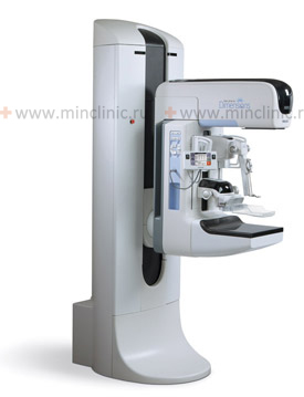
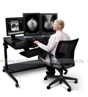
Specialized clinics may use state-of-the-art, full-size digital mammography systems, such as the SELENIA Dimensions system (HOLOGIC Inc., USA), which incorporates 3D tomosynthesis capabilities. These systems are often combined with advanced imaging workstations (e.g., SecureViewDX) for image review and analysis. Furthermore, such high-end equipment can be integrated with horizontal stereotactic systems (e.g., Lorad Multicare Platinum by HOLOGIC Inc.) for precise image-guided breast biopsies. This combination of technology allows for highly efficient breast examinations with an excellent safety profile for the patient due to the use of optimized, minimal doses of X-ray radiation. According to reviews by mammologists, such advanced mammographic diagnostic complexes represent the current best practice in breast imaging.
Benefits of X-ray Digital Mammography with Tomosynthesis
Digital breast tomosynthesis processes the acquired X-ray images to create a series of thin, one-millimeter slices, effectively producing a three-dimensional reconstruction of the breast tissue. This offers several advantages over traditional 2D digital mammography:
- Improved Cancer Detection: Tomosynthesis enables the mammologist to visualize the structure of breast tissue in three dimensions. This helps to reduce the issue of overlapping tissue that can obscure small cancers in 2D mammography, leading to earlier detection of invasive cancers.
- Reduced Recall Rates: By providing clearer images and minimizing tissue overlap, tomosynthesis can reduce the number of women called back for additional imaging due to unclear findings, thereby decreasing patient anxiety and unnecessary biopsies.
- Better Visualization in Dense Breasts: It is particularly beneficial for women with dense breast tissue, where cancers can be harder to detect on 2D mammography.
- Assessment After Implantation: Offers mammologists or plastic surgeons the ability to better assess the condition of the mammary gland after breast augmentation or reconstruction with implants, evaluating implant integrity or identifying issues.
- Enhanced Ductography: When used in conjunction with ductography, tomosynthesis can increase the informational content of the study for evaluating intraductal pathology. The procedure itself is generally well-tolerated by women with any breast size and is typically painless beyond the usual compression.
- Screening and Diagnostic Utility: Tomosynthesis is used both as a screening method for asymptomatic women and for diagnostic evaluation of symptomatic patients.
- Low Radiation Dose: Modern systems deliver a low dose of X-ray radiation, comparable to or only slightly higher than 2D digital mammography, while providing significantly more information.
- High Sensitivity and Information Content: It is a highly sensitive and informative diagnostic method.
- Telemedicine Capabilities: Digital images can be easily transmitted via telecommunication networks for remote consultation by leading mammology specialists from other institutions or countries.
- Shorter Examination Time (for acquisition): The image acquisition phase for tomosynthesis is relatively quick, contributing to efficient patient throughput.
The Optimal Period for a Woman to Have a Breast Mammogram
For premenopausal women, mammographic examination of the breast is ideally recommended to be performed during the early part of the menstrual cycle, typically from day 5 to day 12, counting from the first day of menstruation. During this follicular phase, the mammary glands are generally less tense and softer due to lower estrogen and progesterone levels. Performing mammography during this period makes the examination not only less sensitive or uncomfortable for the woman but also often more informative for the radiologist, as breast tissue is less dense and glandular, allowing for clearer images and better detection of subtle abnormalities.
For postmenopausal women, or those who have had a hysterectomy, mammography can be scheduled at any time.
Indications and Contraindications for Breast Mammography in Women
Indications for Mammography:
- Screening: Regular screening mammography is advisable for asymptomatic women to detect breast cancer at an early, more treatable stage. General guidelines often recommend:
- For women aged 40-49: Discussion with a doctor about individual risk factors and benefits. Some organizations recommend starting annual or biennial screening.
- For women aged 50-74: Annual or biennial (every 1-2 years) screening.
- For women aged 35-40: Screening mammography is typically recommended for those at high risk (e.g., strong family history, known genetic mutations like BRCA1/2). All women after 35-40 years may consider a baseline mammogram, with subsequent frequency determined by risk and guidelines.
- Diagnostic: If a woman experiences any breast symptoms or has abnormal findings on clinical breast examination or other imaging, diagnostic mammography is performed. Symptoms warranting investigation include:
- A new lump or mass in the breast or armpit.
- Breast pain (mastalgia), especially if focal and persistent.
- Nipple discharge (especially if spontaneous, bloody, or unilateral).
- Skin changes on the breast (dimpling, puckering, redness, scaling).
- Nipple changes (retraction, inversion, eczema-like changes).
- Changes in breast size or shape.
- Follow-up: After treatment for breast cancer or for monitoring known benign lesions.
If anything concerning is noted, consultation with a doctor (gynecologist, mammologist, surgeon, or oncologist) is crucial. The specialist will determine the need for mammography and order the appropriate study.
Video explaining the mammography procedure and its importance.
Contraindications for Breast Mammography:
- The only absolute contraindication to breast mammography is pregnancy, due to the potential risk of radiation exposure to the fetus. If breast imaging is essential during pregnancy, ultrasound or MRI (without gadolinium unless absolutely necessary) are preferred.
- Lactation (Breastfeeding): While not an absolute contraindication, mammography during lactation can be less sensitive due to increased breast density. It is often deferred if possible, or performed with special considerations if urgent. Ultrasound is frequently used as a first-line imaging modality in lactating women.
How is the Preparation and Examination of Breast Mammography Conducted?
Preparation:
- On the day of the exam, patients are typically advised not to wear deodorant, talcum powder, or lotion under their arms or on their breasts, as these substances can appear as calcium spots on the mammogram.
- Inform the technologist if there is any possibility of pregnancy, if breastfeeding, if you have breast implants, or if you have any breast symptoms or concerns.
- It's helpful to wear a two-piece outfit for ease of undressing from the waist up.
Examination:
In the department of radiation diagnostics, within the mammography room, the patient is asked to stand or sit in front of the mammography machine. A specially trained radiologic technologist will position one breast at a time between two flat plastic plates (compression paddle and image receptor plate). These plates will compress the breast for a few seconds. Breast compression is necessary to:
- Spread out the breast tissue so that small abnormalities are less likely to be obscured by overlying tissue.
- Reduce the thickness of the breast, allowing for a lower X-ray dose.
- Hold the breast still to prevent blurring of the image due to motion.
A woman typically experiences a sensation of pressure or light discomfort during the compression, which lasts only for a few seconds while the image is taken. A standard screening mammogram usually involves taking at least two X-ray images of each breast from different angles: typically a craniocaudal (CC) view (top-to-bottom) and a mediolateral oblique (MLO) view (side view, angled). For diagnostic mammograms, additional views or spot compression/magnification views may be taken.
After the mammography images are acquired, they are reviewed and interpreted by a qualified radiologist, who specializes in reading medical images, particularly mammograms.
Targeted fine-needle aspiration biopsy (FNAB) or core needle biopsy of breast tissue can be performed using advanced systems like a pistol-needle device guided by stereotactic mammography or ultrasound. This procedure typically involves two stages: imaging localization and precise needle placement for sample acquisition.
Ductography (Galactography) of the Breast Ducts
Ductography, also known as galactography, is a specialized contrast-enhanced X-ray mammography examination specifically designed to visualize the internal structure of the breast ducts. This procedure is indicated for patients who present with pathological nipple discharge, particularly if it is spontaneous, persistent, unilateral, and bloody (sanguineous) or serosanguineous (blood-tinged clear fluid). Ductography is performed to diagnose intraductal abnormalities such as:
- Intraductal papillomas (the most common cause of pathological nipple discharge)
- Duct ectasia
- Fibrocystic changes involving the ducts
- Intraductal carcinomas or ductal carcinoma in situ (DCIS)
Before the ductography procedure, the discharging duct orifice on the nipple is identified. After local anesthesia of the areolar region (if needed, though often not required), a very fine, blunt-tipped cannula is gently inserted into the opening of the specific duct. A small amount of water-soluble iodinated contrast agent is then slowly injected into the duct system. Following contrast injection, standard mammographic X-rays are taken, typically in craniocaudal (CC) and mediolateral oblique (MLO) or true lateral projections. The contrast-enhanced images (ductograms) clearly outline the opacified duct and its branches. The presence of an intraductal breast tumor or other filling defect, ductal narrowing, or distortion will determine its location, size, and characteristics.
Ultrasound Examination of the Breast (Sonography)
Breast ultrasound (sonography) is a valuable imaging modality that uses high-frequency sound waves to create images of the internal structures of the breast. It is often used as an adjunct to mammography or as a primary imaging tool in certain situations. Ultrasound is a simple, widely available, and painless way to detect and characterize breast lesions without using ionizing radiation.
Key applications of breast ultrasound include:
- Differentiating Cysts from Solid Masses: Ultrasound is particularly informative in distinguishing fluid-filled cysts (which are almost always benign) from solid masses, a distinction that can be difficult on mammography alone, especially in dense breasts.
- Evaluating Palpable Lumps: It is often the first imaging test for women under 30-35 with a palpable breast lump (as mammography is less sensitive in young, dense breasts and to avoid radiation).
- Further Evaluation of Mammographic Abnormalities: Used to clarify findings seen on a mammogram, such as focal asymmetries, architectural distortions, or indeterminate masses.
- Imaging Dense Breasts: As a supplemental screening tool in women with dense breast tissue, where mammography sensitivity is reduced.
- Guidance for Interventional Procedures: Ultrasound is commonly used to guide breast biopsies (fine-needle aspiration, core needle biopsy), cyst aspirations, and marker placement.
- Evaluation of Breast Implants.
- Assessing Axillary Lymph Nodes.
Specialized clinics may use expert-class ultrasound devices (e.g., MyLab 70 from Esaote) equipped with an optimal set of functions, high-frequency transducers, and various types of sensitive Doppler imaging (to assess blood flow within lesions). Modern technologies and the versatility of such devices provide the capability for physicians to perform any type of breast biopsy under precise ultrasound guidance.
Magnetic Resonance Imaging (MRI) of the Breast
Breast Magnetic Resonance Imaging (MRI) is a highly sensitive imaging technique that uses strong magnetic fields, radio waves, and a computer to create detailed cross-sectional images of the breast. It is typically performed with the intravenous injection of a gadolinium-based contrast agent.
Breast MRI is not used for routine screening in the general population but plays a crucial role in specific clinical scenarios:
- High-Risk Screening: For women at very high risk of breast cancer (e.g., due to known BRCA gene mutations, strong family history, prior chest radiation), MRI is often recommended as an adjunct to mammography for screening.
- Evaluating Extent of Disease in Newly Diagnosed Breast Cancer: To assess for multifocal or multicentric disease (cancer in more than one quadrant of the breast) or contralateral (opposite) breast cancer.
- Problem Solving: If there are difficulties in interpreting mammography and ultrasound data, or if there is a discrepancy between clinical findings and other imaging (e.g., palpable lump not seen on mammogram/ultrasound).
- Monitoring Response to Neoadjuvant Chemotherapy: To assess how well a tumor is shrinking before surgery.
- Evaluating Breast Implants: MRI is the main method of choice for monitoring the condition of breast implants after cosmetic or reconstructive mammoplasty (prosthetics), particularly for detecting implant rupture (intracapsular or extracapsular).
- Occult Breast Cancer: When cancer is found in an axillary lymph node, but no tumor is evident on mammography or ultrasound, MRI may help locate the primary breast tumor.
- Follow-up of Women with a High Risk of Breast Cancer: MRI is often prescribed for surveillance in this population.
Specialized breast MRI requires dedicated equipment, including an ultra-high-field MR system (e.g., Magnetom Verio from SIEMENS with a magnetic field strength of 3 Tesla) equipped with a special high-tech breast MR coil. The high magnetic field strength of such tomographs provides unprecedented informative value for specialists examining patients with breast diseases. However, MRI has a higher rate of false positives than mammography, often leading to more biopsies.
Magnetic Resonance Imaging (MRI) of the breast is a highly effective tool for evaluating breast implant integrity, capable of detecting issues such as implant rupture or leakage.
Breast Biopsy: Obtaining Tissue for Diagnosis
When imaging studies detect a suspicious abnormality, a breast biopsy is often necessary to obtain a tissue sample for histopathological examination by a pathologist. This is the only way to definitively determine if a lesion is benign or malignant and to establish its specific type.
Types of Biopsy Procedures
Various biopsy techniques are available, chosen based on the lesion's characteristics, location, and visibility on imaging:
- Fine-Needle Aspiration (FNA) Biopsy: A very thin needle is inserted into the lesion to withdraw cells for cytological examination. Often used for palpable lumps or cysts.
- Core Needle Biopsy (CNB): A larger, hollow needle is used to obtain small cores of tissue. This provides more tissue than FNA, allowing for histological examination and receptor testing if cancer is found. CNB is commonly performed under imaging guidance (ultrasound, stereotactic mammography, or MRI).
- Vacuum-Assisted Biopsy (VAB): A type of core biopsy that uses vacuum suction to collect multiple, larger tissue samples through a single needle insertion. Often used for microcalcifications or small lesions.
- Surgical Biopsy (Excisional or Incisional): Involves surgically removing all (excisional) or part (incisional) of the suspicious lesion. This is typically done in an operating room.
Stereotactic Breast Biopsy
This diagnostic procedure can be performed in specialized clinics equipped with systems like the Lorad Multicare Platinum horizontal stereotactic system for breast biopsy, integrated with targeted digital mammography (manufactured by HOLOGIC Inc., USA). This technique is particularly useful for biopsying non-palpable lesions or microcalcifications seen only on mammography.
A targeted fine-needle aspiration biopsy or, more commonly, a core needle biopsy of breast tissue using a pistol-needle system under stereotactic guidance typically consists of two main stages:
- Localization: At the beginning of the biopsy procedure, two sighting mammographic images of the breast area of interest are taken from slightly different angles (stereo pair) using a special technique.
- Targeting and Sampling: With the help of computer processing of the acquired information, a virtual marking of the breast lesion is made, and its precise 3D coordinates are calculated. The length of the biopsy needle is selected. This information is transmitted to the stereotactic device, which then guides the biopsy needle (often part of a pistol-needle system) to the target with an accuracy of approximately 1 mm. The notch in the core biopsy needle allows for the acquisition of breast material sufficient not only for cytological but also for detailed histological examination. This enables crucial ancillary studies if cancer is found, such as immunohistochemical analysis for hormone receptors (estrogen, progesterone), HER2 status, and other tissue prognostic factors.
Differential Diagnosis of Common Breast Findings
Various breast conditions can present with lumps, pain, or imaging abnormalities. A multimodality diagnostic approach helps differentiate them:
| Finding/Symptom | Common Benign Causes | Common Malignant Causes | Key Diagnostic Pointers |
|---|---|---|---|
| Palpable Lump/Mass | Fibroadenoma, Cyst, Fibrocystic changes, Lipoma, Fat necrosis, Duct ectasia, Abscess/Mastitis | Invasive Ductal Carcinoma (IDC), Invasive Lobular Carcinoma (ILC), Medullary carcinoma, Mucinous carcinoma, Phyllodes tumor (can be benign, borderline, or malignant) | Age, characteristics on ultrasound (cystic vs. solid, margins), mammographic appearance, biopsy results. |
| Nipple Discharge | Duct ectasia, Intraductal papilloma, Fibrocystic changes, Pregnancy/Lactation, Medications | Ductal Carcinoma In Situ (DCIS), Invasive Ductal Carcinoma, Paget's disease of the nipple | Character of discharge (bloody, serous, milky, purulent), unilateral vs. bilateral, spontaneous vs. expressed. Ductography, biopsy. |
| Breast Pain (Mastalgia) | Cyclical hormonal changes, Fibrocystic changes, Cysts, Mastitis, Costochondritis, Trauma | Inflammatory Breast Cancer (often diffuse pain, redness, swelling), Large tumors causing stretching | Cyclicality, focality, associated signs of inflammation or mass. Pain is a less common presenting symptom of cancer. |
| Skin Changes (Redness, Dimpling, Peau d'orange) | Mastitis/Abscess, Fat necrosis, Dermatitis | Inflammatory Breast Cancer, Locally advanced breast cancer | Rapid onset, signs of infection vs. progressive skin changes. Biopsy often needed. |
| Microcalcifications on Mammogram | Benign (e.g., fibrocystic changes, duct ectasia, resolving hematoma, skin calcifications) | Ductal Carcinoma In Situ (DCIS), Invasive cancer associated with DCIS | Morphology and distribution of calcifications (e.g., pleomorphic, linear, segmental vs. benign punctate, coarse). Stereotactic biopsy. |
Importance of Multimodality Imaging and Specialist Consultation
No single diagnostic method is perfect for all breast conditions or all women. A combination of clinical examination, mammography, ultrasound, MRI, and biopsy, as indicated, forms a comprehensive "triple assessment" or multimodality approach. This strategy maximizes diagnostic accuracy, allows for early detection of breast cancer, helps characterize benign conditions, and guides appropriate patient management. Consultation with breast specialists (mammologists, radiologists specializing in breast imaging, breast surgeons, oncologists) is crucial for interpreting complex findings and developing individualized care plans.
References
- American College of Radiology (ACR). ACR Practice Parameter for the Performance of Screening and Diagnostic Mammography. Revised 2022.
- Skaane P, Bandos AI, Gullien R, et al. Comparison of digital mammography alone and digital mammography plus tomosynthesis in a population-based screening program. Radiology. 2013 Apr;267(1):47-56.
- Mann RM, Kuhl CK, Kinkel K, Boetes C. Breast MRI: guidelines from the European Society of Breast Imaging. Eur Radiol. 2008 Jul;18(7):1307-18.
- American Cancer Society. American Cancer Society Guidelines for the Early Detection of Cancer. Updated 2023.
- Harvey JA, Mahoney MC, Newell MS, et al. ACR Appropriateness Criteria® Breast Imaging of Pregnant and Lactating Women. J Am Coll Radiol. 2018 May;15(5S):S12-S22.
- Berg WA, Bandos AI, Mendelson EB, et al. Ultrasound as the Primary Screening Test for Breast Cancer: Analysis from ACRIN 6666. J Natl Cancer Inst. 2016 Jan 12;108(4):djv367.
- Parker SH, Burbank F, Jackman RJ, et al. Percutaneous large-core breast biopsy: a multi-institutional study. Radiology. 1994 Aug;193(2):359-64.
- Liberman L. Percutaneous imaging-guided core breast biopsy. AJR Am J Roentgenol. 2000 Jul;175(1):53-61.


