Magnetic Resonance Imaging (MRI) Study Principle
- Understanding Magnetic Resonance Imaging (MRI) Examination
- The Magnetic Resonance Imaging Procedure
- Patient Preparation for an MRI Procedure
- Patient Experience and Tolerability of MRI
- General Indications for Magnetic Resonance Imaging
- Potential Risks, Complications, and Limitations of MRI
- Interpreting MRI Results
- Comparison: MRI vs. Computed Tomography (CT)
- References
Understanding Magnetic Resonance Imaging (MRI) Examination
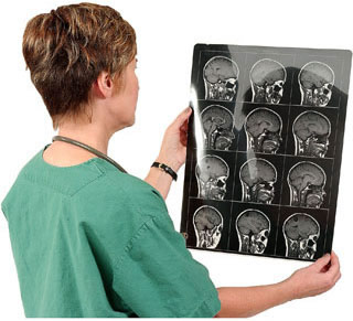
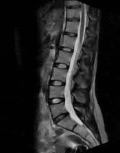
Magnetic Resonance Imaging (MRI) is an advanced medical imaging technique that utilizes a powerful magnetic field, radiofrequency waves, and sophisticated computer technology to produce detailed cross-sectional images of internal body structures. It is widely used to visualize organs such as the brain, cerebral vessels, spine and spinal cord, joints, heart, as well as abdominal and pelvic organs, providing invaluable information for diagnosis and treatment planning. A key advantage of MRI is that, unlike X-rays or Computed Tomography (CT) scans, it does not involve the use of ionizing radiation, meaning the patient does not receive radiation exposure during the diagnostic procedure.
The advent of MRI scanning has had a revolutionary impact on medical practice, particularly in neurology and neurosurgery, comparable to the impact of CT in the 1970s, by offering superior soft tissue contrast and multiplanar imaging capabilities.
Basic Principle of MRI
The technique of magnetic resonance imaging is based on the fundamental principle that tiny particles, specifically hydrogen protons (which are abundant in the water and fat molecules within body tissues), behave like small magnets. When a patient is placed within the strong magnetic field of an MRI scanner, these protons align with the field. A radiofrequency (RF) pulse is then briefly emitted by the scanner, which temporarily knocks these aligned protons out of their equilibrium state. When the RF pulse is turned off, the protons relax back to their original alignment, releasing energy in the form of radio signals. These signals are detected by the MRI scanner's receivers. Different tissues (e.g., gray matter, white matter, CSF, fat, muscle, tumors) have different proton densities and relaxation properties (T1 and T2 relaxation times), causing them to emit signals of varying strengths and at different rates. A powerful computer processes these signals to construct detailed images of the body's internal structures.
Technological Advancements and Scanner Strength
Most commonly, clinical MRI machines utilize a static magnetic field with a strength typically ranging from 0.15 Tesla (T) to 3.0 T. Each Tesla is equivalent to 10,000 Gauss (G); for comparison, the Earth's natural magnetic field strength is approximately 0.5-0.6 G. The radiofrequency field used in MRI is not static; it rotates in a projection perpendicular to the main magnetic field and the patient's body at a speed equal to the Larmor frequency of the targeted protons.
Advanced imaging facilities may offer MRI scans using high-field devices, such as those with a magnetic field strength of 3.0 T. Higher field strengths generally provide better image quality, higher resolution, and faster scan times for certain applications. It is also possible to conduct MRI examinations with intravenous contrast agents (e.g., gadolinium-based Omniscan, though specific agents vary) to enhance the visual difference between healthy tissue and pathological areas like tumors or inflammation. Practical considerations include patient weight restrictions, which for many scanners can be up to 200 kg (approximately 440 lbs).
The Magnetic Resonance Imaging Procedure
The MRI procedure involves several steps to ensure patient safety and optimal image acquisition.
Role of Contrast Agents
Some types of MRI studies require the administration of a special contrast agent to improve the visibility of certain tissues or pathologies. This contrast material is typically gadolinium-based and is administered intravenously (into a vein, usually in the arm or hand). Gadolinium is a paramagnetic substance, meaning its properties enhance the signal intensity of tissues where it accumulates during an MRI scan. Intravenous administration of the contrast agent causes an increase in the signal from tissue sites that have altered vascularity or a disrupted blood-brain barrier as a result of a pathological process (e.g., tumors, inflammation, infection). This provides greater informational content in the obtained images compared to MRI data from the same organ without contrast enhancement, allowing radiologists to better characterize lesions and assess their activity.
Scan Duration and Monitoring
The radiologist and MRI technologist are situated in a separate control room during the MRI procedure. They observe the operation of the scanner and monitor the patient's condition through a large window and via an intercom system. The duration of an MRI study typically ranges from 30 to 60 minutes, but can be longer depending on the anatomical area being examined, the number of sequences required, and whether contrast is used. The patient must remain as still as possible during image acquisition to prevent blurring.
Specific MRI Applications by Body Region
MRI is a versatile tool used to image virtually any part of the body. More information on specific types of magnetic resonance imaging by department and organ can be found via these links:
- General Overview of Magnetic Resonance Imaging (MRI)
- Magnetic Resonance Angiography (MRA) of the Cerebral Vessels
- Magnetic Resonance Imaging (MRI) of the Abdomen
- Magnetic Resonance Imaging (MRI) of the Brain
- Magnetic Resonance Imaging (MRI) of the Cervical Spine
- Magnetic Resonance Imaging (MRI) of the Hip Joint
- Magnetic Resonance Imaging (MRI) of the Knee Joint
- Magnetic Resonance Imaging (MRI) of the Lumbar Spine
- Magnetic Resonance Imaging (MRI) of the Pelvic Organs
- Magnetic Resonance Imaging (MRI) of the Pituitary Gland (Hypophysis)
- Magnetic Resonance Imaging (MRI) of the Shoulder Joint
- Magnetic Resonance Imaging (MRI) of the Thoracic Cavity Organs (Chest MRI - distinct from Thoracic Spine)
- Magnetic Resonance Imaging (MRI) of the Thoracic Spine
- Magnetic Resonance Imaging (MRI) Study Principle (Current Page)
- Whole-Body Magnetic Resonance Imaging (MRI)
Patient Preparation for the Magnetic Resonance Imaging Procedure
General Guidelines
Before a scheduled MRI examination, patients are typically advised not to eat or drink fluids for 4-6 hours prior to the start of the procedure, especially if contrast administration or sedation is anticipated. However, for many standard MRI scans without contrast, fasting may not be strictly necessary; patients should follow the specific instructions provided by the imaging facility.
Screening for Contraindications (Metal Implants)
Prior to an MRI, it is absolutely crucial for patients to inform the medical staff about any metal implants or devices within their body. MRI is generally not possible or not safe if the patient has certain types of metallic implants. These include:
- Clips used for cerebral aneurysms (some older types are ferromagnetic).
- Certain intravascular stents (though many modern stents are MRI-conditional).
- Artificial metal heart valves (some older models).
- Cardiac defibrillators and pacemakers (unless specifically designated as MRI-conditional and protocols for safe scanning are followed).
- Neurostimulators for the brain or spinal cord.
- Cochlear implants in the inner ear.
- Artificial joints (most modern joint prostheses are made of MRI-compatible materials like titanium or cobalt-chromium, but it's essential to confirm).
- Metal plates and screws used for osteosynthesis (bone fixation) – many are MRI-safe or conditional, but older stainless steel implants may cause issues.
- Metal fragments in the body from accidents, explosions, injuries (e.g., shrapnel, metallic foreign bodies in the eyes).
The presence of ferromagnetic metal can lead to blurred or distorted images due to artifacts, and more seriously, poses a risk of displacement (migration) or heating of the implants within the body due to the powerful magnetic field. Recently, manufacturers of medical devices have increasingly begun to produce implantable components using special non-ferromagnetic alloys (e.g., titanium, nitinol) that are not significantly affected by the MRI machine's magnetic field. The manufacturer specifies the product's compatibility with MRI (e.g., "MRI Safe," "MRI Conditional," "MRI Unsafe") in the accompanying documentation, which should always be verified.
Patients should also report any history of kidney disease or dialysis if intravenous contrast is planned, due to the risk of nephrogenic systemic fibrosis (NSF) with certain gadolinium-based contrast agents in individuals with impaired renal function.
Before entering the MRI room, which houses a strong magnetic field (e.g., 3 Tesla), all external metallic objects must be removed to prevent them from becoming projectiles or interfering with the scan. These include:
- Pens, pocket knives, eyeglasses with metal frames.
- Metal jewelry (rings, necklaces, earrings, watches).
- Bank credit cards and security passes (magnetic strips can be erased).
- Hearing aids.
- Metal hairpins, safety pins, clasps, and zippers on clothing.
- Removable dentures with metal components (should be removed before the scan).
Managing Claustrophobia
It is important to clarify in advance if the patient sent for an MRI examination experiences claustrophobia (fear of enclosed spaces). In such cases, several strategies can be employed:
- The patient may need to take a sedative medication prescribed by their doctor beforehand, which can induce slight drowsiness and relieve anxiety.
- Some imaging centers have "open" type MRI machines ("open MRI" or "open-bore MRI"), which have more free space on the sides and a less confining design, often reducing patient anxiety. MRI units with a wider bore (e.g., 70 cm aperture) also tend to be better tolerated by claustrophobic or larger patients, potentially decreasing the need for preliminary sedation.
- Techniques like covering the eyes, listening to music, or having a companion in the room (if permitted and screened for safety) can also help.
Patient Experience and Tolerability of the Magnetic Resonance Imaging Procedure
The MRI procedure itself does not cause any physical pain or direct discomfort to the patient. The main sensations are related to being in an enclosed space and the loud noises produced by the scanner.
In situations where a patient (especially children) cannot lie still or becomes very nervous during the examination, a short-acting sedative drug may be administered to help them calm down and relax throughout the procedure. Any body movements by the patient during the MRI scan will cause motion artifacts, which can blur the acquired images and introduce errors in diagnosis.
The surface of the MRI scanner table may feel cool and hard for some patients. They can often be covered with a blanket for warmth, and a pillow may be placed under their head or knees for comfort, provided this does not interfere with the primary examination area or coil placement.
An intercom system built into the MRI machine allows the patient to communicate with the medical staff (technologist and/or radiologist) located in the adjacent control room throughout the diagnostic procedure. Some MRI scanners are also equipped with built-in video monitors or headphones, allowing the patient to watch a movie or listen to music, which can help distract them and make the experience more tolerable while they are inside the scanner during the image acquisition process.
After the completion of the MRI procedure, the patient can usually resume their normal life and activities immediately. There are typically no restrictions on diet or physical activity, except in cases where sedation was administered. If sedated, the patient will need someone to drive them home and should avoid activities requiring full alertness until the effects of the sedative have worn off.
General Indications for Magnetic Resonance Imaging
MRI allows specialists to obtain detailed images of a patient's organs and tissues—including the brain, cerebral vessels, spine, joints, abdominal organs, pelvic organs, heart, bronchi, and more—to assess their functional state or identify any organic (structural) changes that may have occurred.
MRI is performed in patients in a wide variety of clinical scenarios, including:
- As an alternative to CT angiography (CTA) or conventional angiography to avoid radiation exposure or iodinated contrast risks, particularly for vascular assessment.
- To clarify or further investigate findings from previous imaging studies such as radiography (X-rays) or computed tomography (CT).
- For the diagnosis of pathological tissue growth (tumors, cysts, or other masses) in various organs and soft tissues.
- To assess the state of blood flow and vascular structures (veins, arteries, sinuses, heart) using MRA techniques.
- To evaluate the condition of lymph nodes for enlargement or pathological changes.
- To create multiplanar and three-dimensional reconstructions of organs and anatomical regions from different angles for better spatial understanding.
- For cancer staging: detecting the spread of malignant cells from a primary tumor to other parts of the body (metastases). Determining the stage of cancer development allows physicians to choose the most adequate method of treatment and to make a more accurate prognosis regarding the patient's future condition.
- Evaluation of inflammatory, infectious, traumatic, degenerative, and congenital conditions across various organ systems.
Potential Risks, Complications, and Limitations of Magnetic Resonance Imaging
Safety Regarding Radiation
A significant advantage of MRI is that the machine does not use X-rays (ionizing radiation) in its operation, unlike CT scanners or conventional radiography. To date, there have been no established reports or definitive studies from medical professionals indicating significant, direct harmful side effects on the human body as a result of exposure to the magnetic fields and radio waves used in standard diagnostic MRI procedures when appropriate safety guidelines are followed.
Risks Associated with Contrast Media
Most types of MRI contrast media (contrast agents) contain gadolinium. Intravenous gadolinium-based contrast agents (GBCAs) are generally considered safe for MRI when used appropriately. Allergic reactions to GBCAs administered through a vein are extremely rare in patients, and usually mild if they occur.
However, gadolinium can pose risks for certain patient populations:
- Nephrogenic Systemic Fibrosis (NSF): A rare but serious condition associated with some older GBCAs (linear, non-ionic types) in patients with severely impaired kidney function (e.g., severe chronic kidney disease, patients on dialysis). Newer macrocyclic GBCAs have a much lower risk of NSF. Renal function screening is essential before administering GBCAs to at-risk patients. If a patient has renal insufficiency, caution should be exercised when prescribing MRI studies (e.g., of chest organs or any region) requiring contrast enhancement.
- Gadolinium Deposition: Trace amounts of gadolinium have been found to deposit in the brain and other body tissues after repeated GBCA administrations, even in patients with normal kidney function. The long-term clinical significance of this deposition is still under investigation, but current evidence has not established a direct link to adverse health effects for most patients.
Risks from Magnetic Fields and Metal
The strong magnetic field generated by the MRI machine is the primary source of potential direct risk:
- Damage to Electronic Implants: The magnetic field can damage or cause malfunction of implanted electronic devices such as pacemakers, implantable cardioverter-defibrillators (ICDs), cochlear implants, or certain neurostimulators, unless these devices are specifically certified as "MRI Conditional" and scanned under strict protocols.
- Movement/Displacement of Ferromagnetic Metal: The magnetic field can exert strong forces on ferromagnetic metal fragments (e.g., shrapnel, some older surgical clips) or plates within the body, potentially causing them to move, shift, or heat up, leading to injury.
- Image Artifacts: The presence of metal, even non-ferromagnetic, can cause significant artifacts (distortions) in the MRI images, potentially rendering them non-diagnostic.
- Projectile Effect: Ferromagnetic objects inadvertently brought into the MRI scan room can be forcefully pulled towards the magnet, becoming dangerous projectiles. Strict safety protocols are in place to prevent this.
Practical Limitations
Performing an MRI examination may not always be beneficial or the first choice for several reasons:
- High Cost: MRI examinations are generally more expensive compared to CT tomography or X-rays.
- Long Scan Duration: The MRI scan process is typically longer (30-60 minutes or more) compared to CT scans (often minutes). This can be challenging for patients who have difficulty lying still or are critically ill.
- High Sensitivity to Motion: MRI is very sensitive to movement of internal organs (e.g., breathing, peristalsis) or the whole body during image acquisition, which can degrade image quality more significantly than in CT.
- Claustrophobia: The enclosed nature of most high-field MRI scanners can be intolerable for claustrophobic patients without sedation or use of an open MRI.
- Limited Availability: High-field MRI scanners may not be as widely available as CT scanners in all healthcare settings.
Interpreting MRI Results
If an MRI examination reveals no pathological changes, the study is considered normal. The obtained MRI data, especially in the presence of visible changes, are further analyzed by the patient's attending physician in conjunction with clinical symptoms and other diagnostic findings. This comprehensive assessment allows the physician to develop an appropriate plan for any necessary additional examinations and to formulate an effective treatment strategy.
Comparison: MRI vs. Computed Tomography (CT)
| Feature | Magnetic Resonance Imaging (MRI) | Computed Tomography (CT) |
|---|---|---|
| Imaging Principle | Magnetic fields, radiofrequency waves, proton behavior | X-rays, differential absorption by tissues |
| Ionizing Radiation | No | Yes |
| Soft Tissue Contrast | Excellent, superior for differentiating most soft tissues | Good, but generally less than MRI |
| Bone Detail (Cortical Bone) | Fair to Good (indirectly, by absence of signal) | Excellent |
| Calcification Detection | Poor | Excellent |
| Acute Hemorrhage Detection | Good (especially with specific sequences), but CT often faster and preferred in acute settings | Excellent, very fast for acute intracranial or internal bleeding |
| Scan Time | Longer (typically 20-60+ minutes per body part) | Shorter (typically minutes per body part) |
| Contrast Agents | Gadolinium-based (IV) | Iodinated (IV, Oral, Rectal) |
| Patient Contraindications | Certain metallic implants (pacemakers, some clips, cochlear implants), severe claustrophobia, unstable patients requiring intensive monitoring. | Pregnancy (relative, risk vs. benefit), severe allergy to iodinated contrast, severe renal impairment (for contrast). |
| Common Applications | Neurological imaging (brain, spine), musculoskeletal (joints, soft tissues), abdominal/pelvic soft tissue organs, specialized angiography (MRA), functional imaging. | Trauma (head, spine, body), acute stroke, chest imaging, abdominal/pelvic emergencies, cancer staging, bone imaging, angiography (CTA). |
| Cost | Generally higher | Generally lower |
References
- Westbrook C, Roth C, Talbot J. MRI in Practice. 5th ed. Wiley-Blackwell; 2018.
- Hashemi RH, Bradley WG Jr, Lisanti CJ. MRI: The Basics. 3rd ed. Lippincott Williams & Wilkins; 2010.
- American College of Radiology. ACR Manual on Contrast Media. Version 10.3. 2023.
- Shellock FG. Reference Manual for Magnetic Resonance Safety, Implants and Devices. Biomedical Research Publishing Group; (Updated annually). Access via MRIsafety.com.
- Edelman RR, Hesselink JR, Zlatkin MB, Crues JV. Clinical Magnetic Resonance Imaging. 3rd ed. Saunders Elsevier; 2006.
- Brant WE, Helms CA. Fundamentals of Diagnostic Radiology. 4th ed. Lippincott Williams & Wilkins; 2012.
- Patel PR. Lecture Notes: Radiology. 3rd ed. Wiley-Blackwell; 2010.
- Kanal E, Barkovich AJ, Bell C, et al. ACR guidance document on MR safe practices: 2013. J Magn Reson Imaging. 2013 Mar;37(3):501-30.
See also
- Magnetic Resonance Imaging (MRI)
- Magnetic Resonance Angiography (MRA) of the Cerebral Vessels
- Magnetic Resonance Imaging (MRI) of the Abdomen
- Magnetic Resonance Imaging (MRI) of the Brain
- Magnetic Resonance Imaging (MRI) of the Cervical Spine
- Magnetic Resonance Imaging (MRI) of the Hip Joint
- Magnetic Resonance Imaging (MRI) of the Knee Joint
- Magnetic Resonance Imaging (MRI) of the Lumbar Spine
- Magnetic Resonance Imaging (MRI) of the Pelvic Organs
- Magnetic Resonance Imaging (MRI) of the Pituitary Gland (Hypophysis)
- Magnetic Resonance Imaging (MRI) of the Shoulder Joint
- Magnetic Resonance Imaging (MRI) of the Thoracic Cavity Organs
- Magnetic Resonance Imaging (MRI) of the Thoracic Spine
- Magnetic Resonance Imaging (MRI) Study Principle
- Whole-Body Magnetic Resonance Imaging (MRI)


