Magnetic Resonance Imaging (MRI) of the Pituitary Gland (Hypophysis)
- Understanding MRI of the Pituitary Gland (Hypophysis) and Sella Turcica
- Clinical Indications for Pituitary MRI
- MRI Techniques for Pituitary Imaging
- Patient Preparation and Procedure
- Advantages and Limitations of Pituitary MRI
- Comparison with CT for Pituitary Imaging
- Importance in Diagnosis and Treatment Planning
- References
Understanding Magnetic Resonance Imaging (MRI) of the Pituitary Gland (Hypophysis) and Sella Turcica
Magnetic Resonance Imaging (MRI) of the pituitary gland (also known as the hypophysis) and the sella turcica (the bony cavity at the base of the skull where the pituitary gland is located) is a highly specialized and advanced neuroradiological technique. It is considered one of the most promising and rapidly improving methods for modern diagnostics of this intricate anatomical region.
Role in Neuroradiology
MRI provides unparalleled soft tissue contrast, making it superior to other imaging modalities for visualizing the pituitary gland and its relationship with adjacent structures such as the optic chiasm, cavernous sinuses, and carotid arteries. When conducting an MRI of the pituitary gland and sella turcica, physicians gain the ability to thoroughly investigate subtle structural and pathological changes. This includes evaluating the physicochemical and pathophysiological processes affecting not only the pituitary gland itself but also the surrounding brain structures. Furthermore, MRI allows for functional studies of the brain based on changes in local activity (e.g., fMRI, though less commonly focused solely on the pituitary for function) and can be combined with Magnetic Resonance Angiography (MRA) to assess blood vessels without requiring direct arterial puncture.
Imaging Capabilities
Pituitary MRI utilizes specific protocols with thin slices and high resolution, often including dynamic contrast-enhanced sequences, to optimally visualize the small structures of the pituitary gland and detect subtle abnormalities. It can differentiate the anterior and posterior lobes of the pituitary, the pituitary stalk, and surrounding dural coverings.
Video explaining the MRI procedure for the pituitary gland.
Technology and Contrast Enhancement
For detailed pituitary imaging, high-field MRI scanners, typically 1.5 Tesla (T) or ideally 3.0 T, are preferred as they offer better spatial resolution and signal-to-noise ratio, which is critical for visualizing small lesions like microadenomas. Intravenous administration of a gadolinium-based contrast agent (e.g., Omniscan, though specific agents vary) is almost always performed during a pituitary MRI. The pituitary gland and its stalk normally enhance brightly after contrast due to their rich vascular supply and lack of a typical blood-brain barrier. Pituitary adenomas often show different enhancement patterns (e.g., delayed or less intense enhancement for microadenomas compared to normal gland), which helps in their detection and delineation. Patient weight restrictions (e.g., up to 200 kg) are a practical consideration for the MRI scanner.
Clinical Indications for Pituitary MRI
MRI of the pituitary gland and sella turcica is indicated for a variety of clinical conditions and symptoms suggestive of pituitary or hypothalamic dysfunction or pathology.
Investigating Hyperprolactinemia and Pituitary Tumors
There are numerous reasons for a pathological increase in blood prolactin levels (hyperprolactinemia). One of the primary indications for pituitary MRI is to investigate hyperprolactinemia, as it allows for the visualization of organic changes such as:
- Pituitary Microadenomas: Small tumors (<10 mm in diameter) that are often prolactin-secreting (prolactinomas) and are a common cause of hyperprolactinemia.
- Pituitary Macroadenomas: Larger tumors (≥10 mm in diameter) which can also be prolactinomas or other types of functioning or non-functioning adenomas. Macroadenomas can cause symptoms by compressing adjacent structures like the optic chiasm (leading to visual field defects) or by hormonal overproduction or deficiency.
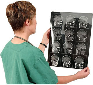
Magnetic Resonance Imaging (MRI) providing a detailed view of the pituitary gland and the sella turcica, crucial for identifying pathologies such as a pituitary adenoma.
Other Common Indications
Beyond hyperprolactinemia, pituitary MRI is indicated for:
- Other Suspected Pituitary Hormone Excess Syndromes: Such as Cushing's disease (ACTH-secreting adenoma), acromegaly/gigantism (GH-secreting adenoma), or TSH-secreting adenomas.
- Hypopituitarism: To investigate causes of pituitary hormone deficiencies.
- Diabetes Insipidus: To evaluate the posterior pituitary and hypothalamus for lesions that might impair ADH production or release.
- Visual Disturbances: Such as bitemporal hemianopsia or other visual field defects, or unexplained optic atrophy, suggestive of optic chiasm compression by a sellar/suprasellar mass.
- Cranial Nerve Palsies: Particularly involving nerves III, IV, V1, V2, and VI, which pass through or near the cavernous sinuses adjacent to the pituitary.
- Headaches: If a pituitary tumor or other sellar pathology is suspected as the cause.
- Empty Sella Syndrome: To diagnose and differentiate primary from secondary empty sella.
- Other Sellar and Parasellar Lesions: Including craniopharyngiomas, Rathke's cleft cysts, meningiomas, germinomas, metastases, inflammatory conditions (hypophysitis), or vascular lesions (aneurysms).
- Monitoring: For follow-up of known pituitary lesions, assessment of treatment response (e.g., after surgery, radiation, or medical therapy for adenomas).
MRI Techniques for Pituitary Imaging
Dedicated pituitary MRI protocols typically include:
- Thin-slice Imaging: Coronal and sagittal T1-weighted images acquired before and after gadolinium contrast administration, with slice thickness usually 3mm or less.
- Dynamic Contrast-Enhanced (DCE) MRI: Involves rapid sequential imaging during and immediately after contrast injection. This is particularly useful for detecting microadenomas, which often enhance differently (usually slower) than the normal pituitary gland.
- T2-Weighted Images: To assess for cystic components, edema, or other pathologies.
- High-Resolution Sequences: Specialized sequences to optimize visualization of the small pituitary gland and surrounding structures.
- Coverage: Imaging usually includes the entire sella turcica, suprasellar cistern, cavernous sinuses, and optic chiasm.
Patient Preparation and Procedure
Preparation for a pituitary MRI is generally minimal:
- Screening: Patients are screened for MRI contraindications (e.g., incompatible metallic implants, severe claustrophobia).
- Metal Objects: All removable metallic items must be taken off.
- Fasting: Usually not required unless sedation is planned.
- Contrast Agent: An IV line will be placed for gadolinium administration. Patients should report any allergies or history of kidney disease.
The procedure involves the patient lying still on the MRI table while it moves into the scanner. The scan time for a dedicated pituitary MRI is typically 30-45 minutes. Earplugs or headphones are provided due to the loud operating noise.
Advantages and Limitations of Pituitary MRI
Advantages:
- Superior soft tissue contrast for visualizing the pituitary gland and surrounding structures.
- No ionizing radiation.
- Multiplanar imaging capability.
- High sensitivity for detecting small pituitary lesions (microadenomas).
- Dynamic contrast enhancement provides functional information about lesion vascularity.
Limitations:
- Susceptibility to motion artifacts.
- Longer scan time compared to CT.
- Higher cost.
- MRI contraindications (metallic implants, claustrophobia).
- Gadolinium contrast risks (though rare with proper screening).
- Difficulty in differentiating very small adenomas from normal pituitary heterogeneity or incidental cysts in some cases.
- Calcifications are better seen on CT.
Comparison with CT for Pituitary Imaging
| Feature | Pituitary MRI | Pituitary CT Scan |
|---|---|---|
| Primary Utility | Detailed evaluation of pituitary gland, sella, parasellar soft tissues, detection of adenomas (especially microadenomas), assessment of optic chiasm/cavernous sinus involvement. | Evaluation of bony sella turcica, detection of calcifications within lesions, acute hemorrhage, or when MRI is contraindicated. Can show larger macroadenomas. |
| Soft Tissue Contrast | Excellent | Fair to Good (with contrast) |
| Detection of Microadenomas (<10mm) | High sensitivity, especially with dynamic contrast. | Lower sensitivity; may miss small lesions. |
| Visualization of Optic Chiasm & Cavernous Sinuses | Excellent | Good, but less detail than MRI. |
| Ionizing Radiation | No | Yes |
| Contrast Agent | Gadolinium-based (IV) - almost always used. | Iodinated (IV) - often used. |
| Scan Time | Longer (30-45 min) | Shorter (5-10 min) |
MRI is generally considered the gold standard for imaging the pituitary gland and sella turcica due to its superior soft tissue resolution and ability to detect subtle lesions.
Importance in Diagnosis and Treatment Planning
MRI of the pituitary gland is indispensable for the accurate diagnosis of a wide range of pituitary and parasellar pathologies. It provides critical information for determining the nature, size, and extent of lesions, their relationship to adjacent vital structures (like the optic chiasm and carotid arteries), and helps guide subsequent management decisions, including whether medical therapy, surgery (e.g., transsphenoidal adenomectomy), or radiation therapy is most appropriate. It is also essential for postoperative follow-up and monitoring treatment response.
References
- Bonneville F, Cattin F, Marsot-Dupuch K, Dormont D, Bonneville JF, Chiras J. T1 signal hyperintensity in the sellar region: spectrum of findings. Radiographics. 2006 Jan-Feb;26(1):93-113.
- Elster AD. Modern imaging of the pituitary. Radiology. 1993 Sep;188(3):609-20.
- Kucharczyk W, Montanera WJ. The sella and parasellar region. In: Atlas SW, ed. Magnetic Resonance Imaging of the Brain and Spine. 4th ed. Lippincott Williams & Wilkins; 2009:chap 18.
- Osborn AG, Salzman KL, Katzman G, et al. Diagnostic Imaging: Brain. 2nd ed. Amirsys; 2009.
- Molitch ME. Diagnosis and treatment of pituitary adenomas: a review. JAMA. 2017 May 16;317(5):516-524.
- Katznelson L, Laws ER Jr, Melmed S, et al. Acromegaly: an endocrine society clinical practice guideline. J Clin Endocrinol Metab. 2014 Nov;99(11):3933-51. (Context for pituitary adenoma imaging).
- Faje A. Cushing's Disease: A Multidisciplinary Approach to Diagnosis and Management. J Clin Endocrinol Metab. 2018 Jul 1;103(7):2469-2481.
- American College of Radiology. ACR Appropriateness Criteria® Pituitary Adenoma. Last review date: 2021.
See also
- Magnetic Resonance Imaging (MRI)
- Magnetic Resonance Angiography (MRA) of the Cerebral Vessels
- Magnetic Resonance Imaging (MRI) of the Abdomen
- Magnetic Resonance Imaging (MRI) of the Brain
- Magnetic Resonance Imaging (MRI) of the Cervical Spine
- Magnetic Resonance Imaging (MRI) of the Hip Joint
- Magnetic Resonance Imaging (MRI) of the Knee Joint
- Magnetic Resonance Imaging (MRI) of the Lumbar Spine
- Magnetic Resonance Imaging (MRI) of the Pelvic Organs
- Magnetic Resonance Imaging (MRI) of the Pituitary Gland (Hypophysis)
- Magnetic Resonance Imaging (MRI) of the Shoulder Joint
- Magnetic Resonance Imaging (MRI) of the Thoracic Cavity Organs
- Magnetic Resonance Imaging (MRI) of the Thoracic Spine
- Magnetic Resonance Imaging (MRI) Study Principle
- Whole-Body Magnetic Resonance Imaging (MRI)


