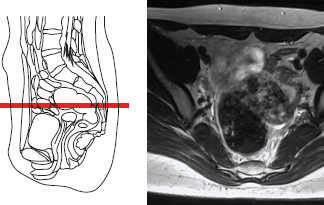
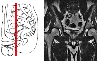
Magnetic Resonance Imaging (MRI) of the Pelvic Organs
- Understanding MRI of the Pelvic Organs
- Indications for Pelvic MRI
- Common Pathological Processes Diagnosed by Pelvic MRI
- Patient Preparation for Pelvic MRI
- The Pelvic MRI Procedure
- MRI Techniques for Pelvic Imaging and Contrast Use
- Advantages and Limitations of Pelvic MRI
- Comparison with Other Pelvic Imaging Modalities
- Related Diseases of the Genitourinary System
- References
Understanding Magnetic Resonance Imaging (MRI) of the Pelvic Organs
Magnetic Resonance Imaging (MRI) of the pelvic organs is a highly advanced, non-invasive diagnostic imaging technique that utilizes strong magnetic fields, radiofrequency waves, and computer processing to create detailed cross-sectional images of the organs and soft tissues within the pelvis. This includes structures such as the bladder, prostate gland, seminal vesicles in males, and the uterus, ovaries, and fallopian tubes in females, as well as the rectum, pelvic muscles, lymph nodes, and blood vessels.
Advantages of Pelvic MRI
The primary advantages of pelvic MRI include:
- Incredibly High Diagnostic Efficiency: MRI often provides superior soft tissue contrast and detail compared to other clinical diagnostic tests like ultrasound or CT for many pelvic conditions.
- Harmlessness of the Examination (No Ionizing Radiation): Unlike CT scans and X-ray diagnostics, MRI does not use ionizing radiation, making it a safer option for repeated examinations or for patients sensitive to radiation.
- High Resolution: MRI of the pelvic organs can visualize objects and anatomical details down to several millimeters in size. It also offers the capability to obtain images in any desired plane (axial, sagittal, coronal, or oblique) without repositioning the patient.
- Comprehensive Tissue Visualization: MRI can effectively visualize a wide range of tissues under normal conditions and in the presence of various pathologies, including inflammation, tumors, and congenital anomalies.
Video explaining the MRI procedure for pelvic organs.
Role of Preliminary Ultrasound
MRI of the pelvic organs is often performed after a preliminary ultrasound examination (transabdominal or transvaginal/transrectal) of this anatomical region. Ultrasound serves as a valuable initial screening tool due to its accessibility and cost-effectiveness. It is advisable for patients to bring previous ultrasound reports and images to their MRI appointment, as this comparative information can enhance the diagnostic interpretation of the MRI findings, even if the ultrasound was reported as normal.
Technology and Scanner Strength
For optimal pelvic imaging, MRI studies are best performed on scanners with a magnetic field strength of at least 1.5 Tesla (T). High-field MRI, such as 3.0 T systems, can offer even greater detail and faster scan times for certain sequences. Intravenous contrast agents (e.g., gadolinium-based, like Omniscan, though specific agents vary) may be administered to enhance the visualization of vascular structures, inflammation, or to better characterize tumors by increasing the visual difference between healthy and pathological tissue. Patient weight restrictions (e.g., up to 200 kg) apply to most MRI scanners.
Indications for Pelvic MRI
Indications for performing an MRI of the pelvic organs in both men and women arise in various clinical situations:
General Indications for Men and Women
- Traumatic Pelvic Injuries: To assess soft tissue damage, hematomas, or occult fractures.
- Suspicion of Tumors: Evaluation of suspected tumors of the bladder, rectum, or other pelvic soft tissue structures.
- Staging of Known Pelvic Tumors: To assess the local extent of tumors and their spread to adjacent structures or regional lymph nodes.
- Inflammatory Conditions: Such as vesiculitis (inflammation of seminal vesicles), proctitis (inflammation of the rectum), or pelvic inflammatory disease (PID) in women.
- Chronic Pelvic Pain: When the cause is unclear after initial investigations.
- Sacral Pain or Sacrodynia: To evaluate for sacral pathologies, nerve impingement, or soft tissue abnormalities.
- Assessment of Regional Lymph Nodes: For detecting metastatic spread or inflammatory changes.
- Congenital Anomalies of the pelvic organs or urogenital system.
- Vascular Pathologies: Such as pelvic congestion syndrome or arteriovenous malformations.
- Acute Conditions (in select cases): Such as suspected ovarian torsion or complications of appendicitis/diverticulitis involving the pelvis.
Specific Indications for Prostate MRI
For men, a primary indication for pelvic MRI is the evaluation of prostate cancer. MRI plays a crucial role in:
- Local and Regional Staging: Determining the extent of the tumor within the prostate, assessing for extracapsular extension, seminal vesicle invasion, and involvement of regional lymph nodes. The high specificity of prostate MRI (approximately 88–90%) makes it essential for patients with a medium to high risk of extra-organ tumor spread.
- Guiding Treatment Decisions: Helping to determine the appropriateness of surgical treatment (e.g., radical prostatectomy) versus radiation therapy or other modalities.
- Detection of Clinically Significant Cancer: Multiparametric MRI (mpMRI) of the prostate is increasingly used to detect and characterize suspicious lesions, especially in men with elevated prostate-specific antigen (PSA) levels and prior negative biopsies, or for active surveillance.
- Assessment of Regional Lymph Nodes and Bone Metastases: Pelvic MRI can surpass CT in diagnostic accuracy for detecting metastases in pelvic lymph nodes and can also identify bone metastases in the pelvic bones and lumbar spine.
Analysis of decision-making processes in prostate cancer management convincingly proves the necessity of performing an MRI of the prostate in patients with a blood PSA level greater than 10 ng/ml, or with other high-risk features.
Common Pathological Processes Diagnosed by Pelvic MRI
There are several potential reasons for the development of pathology within the pelvic organs. MRI can recognize and differentiate many of these processes:
- Inflammatory Conditions:
- In women: Adnexitis (inflammation of ovaries/fallopian tubes), endometritis (inflammation of uterine lining), pelvic inflammatory disease (PID).
- In men: Prostatitis (inflammation of the prostate), vesiculitis (inflammation of seminal vesicles), orchitis (inflammation of testes).
- In both: Proctitis (inflammation of rectum), cystitis (bladder inflammation).
- Tumors or Other Proliferative Processes:
- In women: Uterine fibroids (leiomyomas), ovarian cysts/tumors, endometrial cancer/polyps, cervical cancer.
- In men: Benign Prostatic Hyperplasia (BPH), prostate cancer.
- In both: Bladder tumors, rectal polyps/cancer, soft tissue sarcomas.
- Diseases Associated with Vascular Pathology: Hemorrhoids, pelvic congestion syndrome.
- Acute Conditions: Ovarian apoplexy (ruptured ovarian cyst/hemorrhage), bladder rupture (traumatic), acute appendicitis with pelvic involvement, diverticulitis complications.
- Other Diseases of the Genitourinary System: Endometriosis, adenomyosis, congenital anomalies.
Patient Preparation for Pelvic MRI
Pelvic MRI often does not require extensive special preparation, but some general guidelines apply, and specific instructions may be given by the imaging center:
- Fasting: For some pelvic MRI studies, especially those evaluating bowel or requiring contrast, fasting for 4-6 hours may be requested to reduce bowel motion and improve image quality.
- Bladder Filling: For both men and women, some (not tight or overly full) filling of the urinary bladder is often required. It is generally recommended not to empty the bladder immediately before the study. A moderately full bladder helps to displace bowel loops and provides a good acoustic window if ultrasound is also performed, and can improve delineation of bladder and adjacent structures on MRI.
- Bowel Preparation (Sometimes): For specific indications like MRI for rectal cancer staging or endometriosis involving the bowel, mild bowel preparation (e.g., clear liquids, laxative) or an enema might be requested to reduce bowel contents and gas. Anti-peristaltic medication (e.g., glucagon or hyoscine butylbromide) may be given just before the scan to reduce bowel motion.
- Metal Objects & Screening: Standard MRI safety screening for metallic implants and removal of all external metallic items is mandatory.
- Menstrual Cycle (for female pelvic MRI for specific conditions): For certain conditions like endometriosis, the MRI may be timed according to the menstrual cycle for optimal visualization, though this is not always necessary.
Patients should always follow the specific preparation instructions provided by the facility performing the MRI.
The Pelvic MRI Procedure
The procedure for a pelvic MRI is similar to MRIs of other body regions:
- The patient changes into a hospital gown and removes all metallic objects.
- An IV line may be placed if contrast is to be used.
- The patient lies on the MRI table, usually supine (on their back). A specialized pelvic surface coil (an antenna-like device) is placed over or around the pelvis to improve image quality.
- The table slides into the MRI scanner tunnel.
- The patient needs to remain very still during the scan, which can last 30-60 minutes or longer, depending on the sequences performed and whether contrast is used. Loud noises are produced by the scanner; earplugs or headphones are provided.
- The technologist communicates with the patient via an intercom.
For some specific applications (e.g., endorectal MRI for prostate or rectal cancer), a small coil may be gently inserted into the rectum to obtain very high-resolution images of the target organ, though this is less common with modern external coils and higher field strength magnets.
MRI Techniques for Pelvic Imaging and Contrast Use
Pelvic MRI protocols utilize a variety of sequences tailored to the specific organs and suspected pathology:
- T1-Weighted Images (T1W): Provide good anatomical detail, useful for identifying fat (e.g., in dermoid cysts) and hemorrhage. Post-contrast T1W images are crucial for assessing vascularity, inflammation, and tumor enhancement.
- T2-Weighted Images (T2W): Excellent for visualizing fluid-filled structures (e.g., bladder, cysts, edema) and delineating zonal anatomy of organs like the prostate and uterus. Pathological processes often appear bright on T2W images. Fat suppression techniques are often used with T2W imaging to make fluid and inflammation more conspicuous.
- Diffusion-Weighted Imaging (DWI): Sensitive for detecting restricted diffusion, which can be seen in malignant tumors, abscesses, and acute ischemia. It helps in characterizing lesions and assessing treatment response.
- Dynamic Contrast-Enhanced (DCE) MRI: Involves acquiring images rapidly before, during, and after intravenous contrast injection. It provides information about tissue vascularity and perfusion, useful for characterizing tumors (e.g., prostate cancer, ovarian tumors) and assessing inflammatory activity.
- Specialized Sequences: Such as MR spectroscopy (for metabolic information in prostate cancer), or sequences optimized for detecting endometriosis implants.
Intravenous gadolinium-based contrast is frequently used in pelvic MRI to improve the detection and characterization of tumors, inflammation, infection, and to assess vascularity.
Advantages and Limitations of Pelvic MRI
Advantages:
- Superior soft tissue contrast compared to CT and ultrasound for many pelvic pathologies.
- No ionizing radiation.
- Multiplanar imaging capability.
- Excellent for staging many pelvic cancers (prostate, rectal, cervical, endometrial).
- Problem-solving tool when other imaging is inconclusive.
- Functional information from DWI, DCE-MRI, and MRS.
Limitations:
- Higher cost and longer scan times compared to ultrasound or CT.
- Susceptibility to motion artifacts from bowel peristalsis or patient movement.
- MRI contraindications (certain metallic implants, severe claustrophobia).
- Less sensitive than CT for detecting calcifications or acute bone trauma.
- Gadolinium contrast risks (though low with proper screening).
- Interpretation requires expertise in pelvic MRI.
Comparison with Other Pelvic Imaging Modalities
| Modality | Primary Strengths for Pelvic Imaging | Primary Weaknesses for Pelvic Imaging |
|---|---|---|
| Pelvic MRI | Excellent soft tissue detail (uterus, ovaries, prostate, rectum, bladder wall), staging pelvic cancers, detecting endometriosis, evaluating complex masses/fistulae. No radiation. | Cost, scan time, motion artifacts, less bone/calcification detail than CT. Some contraindications. |
| Pelvic Ultrasound (Transabdominal/Transvaginal/Transrectal) | Initial evaluation of uterus/ovaries (TVUS), prostate (TRUS), bladder. Detects free fluid, cysts, some masses. Real-time, inexpensive, no radiation, portable. Guiding biopsies. | Operator dependent, limited field of view, bowel gas artifact, less soft tissue detail than MRI, limited for deep pelvic structures or staging extensive disease. |
| Pelvic CT Scan | Detecting calcifications/stones, acute trauma/hemorrhage, bone involvement by tumors, staging some cancers (especially for distant mets), guidance for some biopsies/drainages. Faster than MRI. | Ionizing radiation, poorer soft tissue contrast than MRI for many pelvic conditions. Iodinated contrast risks. |
| PET-CT | Detecting metabolically active tumors, staging/restaging certain cancers, assessing treatment response. | Radiation (from CT and PET tracer), lower anatomical resolution than MRI/CT alone, cost, availability. |
Related Diseases of the Urinary-Reproductive System
Pelvic MRI is instrumental in diagnosing and managing a wide range of diseases affecting the genitourinary and lower gastrointestinal systems. Some relevant conditions include:
- Benign Prostatic Hyperplasia (BPH)
- Cystitis, Urocystitis (Bladder Inflammation)
- Hydrocele (Fluid around Testicle)
- Kidney Stones (Urolithiasis) - (can cause pelvic pain if in lower ureter/bladder)
- Kidney (Urinary) Syndromes and Urinalysis Findings:
- Bilirubinuria and Urobilinogenuria
- Cylindruria (Casts in the urine)
- Glucosuria (Glucose in the urine)
- Hematuria (Blood in the urine)
- Hemoglobinuria (Hemoglobin in the urine)
- Ketonuria (Ketone bodies in the urine)
- Myoglobinuria (Myoglobin in the urine)
- Proteinuria (Protein in the urine)
- Purpurinuria, Porphyrinuria (Porphyrins in the urine)
- Pyuria, Leukocyturia (WBC in the urine)
- Orchitis, Didymitis (Testicular Inflammation)
- Prostatitis
- Pyelonephritis (Kidney Infection - can cause pelvic discomfort)
- Hydronephrosis, Pyonephrosis (Kidney Swelling/Infection)
- Varicocele
- Vesiculitis (Seminal Vesicle Inflammation)
- Endometriosis, Adenomyosis, Uterine Fibroids, Ovarian Cysts/Tumors.
- Rectal Cancer, Inflammatory Bowel Disease involving the rectum.
References
- Hricak H, Thoeni RF. Magnetic resonance imaging of the pelvis: a practical approach. Edward Arnold; 1993.
- Weinreb JC, Brown J, et al. ACR Appropriateness Criteria® Staging of Prostate Cancer. J Am Coll Radiol. 2020 Nov;17(11S):S552-S563.
- American College of Radiology. ACR Appropriateness Criteria® Pelvic Pain in the Nonpregnant Patient. Last review date: 2019.
- Jha P, Sekhon S, Khan M, et al. MRI of Endometriosis: A Comprehensive Review. Radiographics. 2020 Sep-Oct;40(5):1477-1502.
- Kaur H, Silverman PM, Scatarige JC, et al. MR imaging of the uterine cervix. Radiographics. 1993 Jul;13(4):825-42.
- Barentsz JO, Richenberg J, Clements R, et al. ESUR prostate MR guidelines 2012. Eur Radiol. 2012 Apr;22(4):746-57. (Though guidelines evolve, this is a foundational reference).
- Forstner R, Cunha TM, Hamm B. MRI in staging of rectal cancer: an update. Eur Radiol. 2010 Aug;20(8):1791-6.
- Sala E, Rockall AG, Rangarajan D, Kubik-Huch RA. The role of dynamic contrast-enhanced and diffusion weighted magnetic resonance imaging in the female pelvis. Eur J Radiol. 2010 Mar;73(3):497-507.
See also
- Magnetic Resonance Imaging (MRI)
- Magnetic Resonance Angiography (MRA) of the Cerebral Vessels
- Magnetic Resonance Imaging (MRI) of the Abdomen
- Magnetic Resonance Imaging (MRI) of the Brain
- Magnetic Resonance Imaging (MRI) of the Cervical Spine
- Magnetic Resonance Imaging (MRI) of the Hip Joint
- Magnetic Resonance Imaging (MRI) of the Knee Joint
- Magnetic Resonance Imaging (MRI) of the Lumbar Spine
- Magnetic Resonance Imaging (MRI) of the Pelvic Organs
- Magnetic Resonance Imaging (MRI) of the Pituitary Gland (Hypophysis)
- Magnetic Resonance Imaging (MRI) of the Shoulder Joint
- Magnetic Resonance Imaging (MRI) of the Thoracic Cavity Organs
- Magnetic Resonance Imaging (MRI) of the Thoracic Spine
- Magnetic Resonance Imaging (MRI) Study Principle
- Whole-Body Magnetic Resonance Imaging (MRI)


