Magnetic Resonance Imaging (MRI) of the Cervical Spine
- Understanding MRI of the Cervical Spine (Neck)
- Clinical Indications for Cervical Spine MRI
- MRI Techniques and Technology
- Patient Preparation and Procedure
- Advantages and Limitations of Cervical Spine MRI
- Comparison with Other Cervical Spine Imaging Modalities
- Interpreting Cervical Spine MRI: Examples
- The Role of Cervical MRI in Diagnosis and Management
- References
Understanding Magnetic Resonance Imaging (MRI) of the Cervical Spine (Neck)
Magnetic Resonance Imaging (MRI) of the cervical spine (the neck region) is a premier, non-invasive diagnostic imaging modality that provides highly detailed images of the vertebrae, intervertebral discs, spinal cord, nerve roots, and surrounding soft tissues of the neck. It is considered one of the most promising and rapidly advancing methods in modern diagnostics for spinal pathologies.
Diagnostic Capabilities and Principles
By utilizing strong magnetic fields, radiofrequency pulses, and sophisticated computer analysis, cervical spine MRI allows physicians to not only investigate structural and pathological changes but also to assess certain physicochemical and pathophysiological processes affecting the entire cervical spine or its individual components. This detailed visualization is crucial for diagnosing a wide array of conditions causing neck pain, arm pain (radiculopathy), weakness, numbness, or symptoms of spinal cord compression (myelopathy).
Video explaining the cervical spine MRI procedure and what to expect.
Imaging Congenital and Acquired Pathologies
Developmental defects of the spine, while more common in the lumbosacral region, also occur in the cervical spine. MRI can identify congenital malformations such as occipitalization of the atlas (fusion of C1 to the occiput), atlantoaxial instability (mixing of the C1 vertebra in relation to C2), or Klippel-Feil syndrome (short neck syndrome), where cervical vertebrae may appear as a shapeless bone mass. Patients with Klippel-Feil syndrome often present with a visibly short neck (head appearing to lie directly on the body), limited head mobility, a low posterior hairline, and associated scoliosis or kyphoscoliosis.
Accessory cervical ribs, another congenital anomaly, can sometimes cause Accessory Cervical Rib Syndrome (a form of thoracic outlet syndrome) if they impinge on neurovascular structures. Symptoms may be clinically manifested under the influence of adverse factors like cooling, trauma, or infection. In many instances, additional cervical ribs are asymptomatic and discovered as an accidental finding during cervical spine MRI. Clinical manifestations, when present, are characterized by neuralgic pain in the shoulder, which can sometimes extend down the entire upper limb, and may include vascular symptoms.
Beyond congenital issues, MRI excels at detecting acquired pathologies such as traumatic changes, degenerative disc disease, tumors, and inflammatory conditions.
3D Reconstruction and Surgical Planning
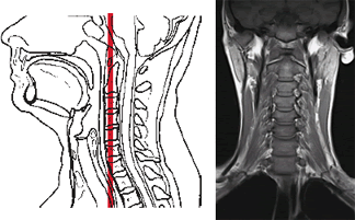
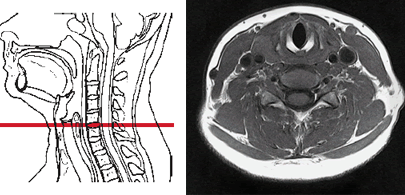
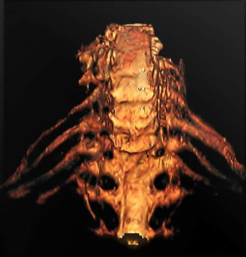
MRI of the cervical spine allows for the acquisition of a series of thin cross-sectional images in multiple planes (sagittal, axial, coronal). These images can be processed to create three-dimensional (3D) reconstructions of the area under study. This advanced visualization can highlight the vascular network (when MRA sequences are included) and even delineate individual nerve trunks and blood vessels passing within the projection of the cervical spine.
Such detailed 3D reconstructions provide invaluable assistance to neurosurgeons and spine surgeons in precisely planning operations on the cervical spine and spinal cord, and for subsequent postoperative monitoring of the patient's spine and assessment of treatment efficacy.
Early diagnosis using MRI of the cervical spine for symptoms like pain or discomfort in the neck and occipital region allows for timely treatment of diseases affecting the cervical spine and spinal cord at this level, potentially preventing progression and long-term disability.
A key advantage of MRI is its ability to simultaneously demonstrate the bony spine itself, the intervertebral discs, ligaments, spinal cord, nerve roots, and surrounding soft tissues (muscles, tendons) over a large area, often without the need for intravenous contrast agents (though contrast is used for specific indications like tumors, infection, or inflammation) and, importantly, without the use of ionizing radiation (as in X-rays or CT scans). MRI can accurately determine the localization and size of tumors, assess the cartilaginous surfaces of facet joints, and evaluate the condition of muscles and tendons.
Currently, MRI of the cervical spine has become the primary imaging modality for diagnosing most diseases of this region, often providing more comprehensive information than radiography or computed tomography (CT).
Clinical Indications for Cervical Spine MRI
An MRI of the cervical spine may be prescribed by a physician in various situations, including:
- Osteochondrosis of the cervical spine (cervical spondylosis): Degenerative changes affecting the discs, vertebral bodies, and facet joints.
- Cervical Radiculopathy: Symptoms of nerve root compression, such as pain, numbness, tingling, or weakness radiating into the shoulder, arm, or hand, often caused by protrusion or herniated discs of the cervical spine or foraminal stenosis.
- Cervical Myelopathy: Symptoms of spinal cord compression, such as gait disturbance, balance problems, weakness or clumsiness in the hands or legs, and bowel/bladder dysfunction.
- Tumors and Metastases: Evaluation of primary tumors of the spine or spinal cord, or detection of metastatic tumor cells at the level of the cervical spine.
- Spinal stenosis: Narrowing of the spinal canal or neural foramina.
- Injuries to the cervical spine: Including fractures, dislocations, ligamentous injuries, or instability of the spine, and assessment of traumatic spinal cord injury.
- Developmental anomalies of the cervical spine: Such as Klippel-Feil syndrome (short neck syndrome), accessory cervical rib syndrome, Chiari malformations, or syringomyelia.
- Inflammatory and Infectious Conditions: Such as discitis, vertebral osteomyelitis, epidural abscess, transverse myelitis, or rheumatological conditions affecting the spine (e.g., rheumatoid arthritis, ankylosing spondylitis).
- Demyelinating Diseases: To detect plaques of Multiple Sclerosis (MS) within the cervical spinal cord.
- Vascular Malformations or Ischemia of the Spinal Cord.
- Preoperative Planning and Postoperative Assessment.
MRI Techniques and Technology
High-field MRI scanners, typically 1.5 Tesla (T) or 3.0 T, are used for cervical spine imaging to achieve optimal image quality and resolution. Various pulse sequences are employed to highlight different tissue characteristics:
- T1-Weighted Images (T1W): Provide excellent anatomical detail of the vertebrae, discs, and spinal cord. Fat appears bright.
- T2-Weighted Images (T2W): Highly sensitive to fluid and pathology. CSF, edema, inflammation, and most lesions appear bright. Discs are well-visualized.
- STIR (Short Tau Inversion Recovery) or Fat-Saturated T2W: Suppresses the signal from fat, making fluid and edema (e.g., bone bruises, inflammation) more conspicuous.
- Gradient Echo (GRE) Sequences: Can be useful for assessing foraminal stenosis and visualizing bony spurs.
- Post-Contrast T1-Weighted Images: Acquired after intravenous administration of a gadolinium-based contrast agent (e.g., Omniscan, though specific agents vary). Contrast enhances areas of inflammation, infection, tumors, or disruption of the blood-spinal cord barrier, increasing the visual difference between healthy tissue and pathology.
Patients may undergo an MRI of the cervical spine using an apparatus with a magnetic field of 3.0 T for superior detail. Patient weight restrictions (e.g., up to 200 kg) are a consideration for the MRI scanner table.
Patient Preparation and Procedure
Preparation for a cervical spine MRI is generally minimal:
- Screening: Patients complete a safety questionnaire to identify any MRI contraindications (e.g., certain pacemakers, metallic implants, severe claustrophobia, pregnancy).
- Metal Objects: All removable ferromagnetic items (jewelry, hairpins, etc.) must be removed.
- Clothing: Patients typically change into a hospital gown.
- Fasting: Usually not required for a standard cervical spine MRI without sedation. If IV contrast or sedation is planned, specific instructions will be provided.
During the procedure, the patient lies supine on the MRI table, often with their head and neck positioned within a specialized surface coil designed for cervical spine imaging. The table then slides into the MRI scanner. The patient must remain very still throughout the scan, which can last from 20 to 45 minutes, depending on the number of sequences performed. The scanner produces loud tapping or knocking sounds, and earplugs or headphones are provided for patient comfort.
Advantages and Limitations of Cervical Spine MRI
Advantages:
- Superior soft tissue contrast for visualizing the spinal cord, nerve roots, intervertebral discs, ligaments, and surrounding soft tissues.
- No ionizing radiation exposure, making it safe for repeated studies.
- Multiplanar imaging capability (sagittal, axial, coronal) without repositioning the patient.
- Highly sensitive for detecting a wide range of pathologies, including disc herniations, spinal cord lesions (tumors, inflammation, demyelination), ligamentous injuries, and infections.
- Can non-invasively assess vascular structures with MRA sequences if needed.
Limitations:
- Less sensitive than CT for detecting acute cortical bone fractures or fine bony detail.
- Longer scan times compared to CT, making it more susceptible to motion artifacts from patient movement or swallowing.
- Higher cost than X-ray or CT.
- Contraindicated in patients with certain incompatible metallic implants.
- Can be challenging for claustrophobic patients or those unable to lie still for the required duration.
- MRI findings (e.g., degenerative changes, disc bulges) are common in asymptomatic individuals, requiring careful clinical correlation.
Comparison with Other Cervical Spine Imaging Modalities
| Modality | Principle | Radiation | Primary Strengths for Cervical Spine | Primary Weaknesses for Cervical Spine |
|---|---|---|---|---|
| Cervical Spine MRI | Magnetic fields, radio waves | No | Spinal cord, nerve roots, discs, ligaments, soft tissues, tumors, infection/inflammation, demyelination. | Longer scan, motion sensitive, less bone detail than CT, cost, MRI contraindications. |
| Cervical Spine CT Scan | X-rays | Yes | Excellent bone detail (fractures, degenerative bony changes, stenosis), calcified discs/ligaments. Faster than MRI, good in trauma. | Radiation, poorer soft tissue/cord detail than MRI. Iodinated contrast risks if used (e.g., for CTA or CT myelography). |
| Cervical Spine X-ray | X-rays | Yes (lower than CT) | Initial assessment of alignment (flexion/extension views for instability), fractures, gross degenerative changes, bone lesions. Inexpensive, widely available. | Limited soft tissue/cord/disc detail. Superimposition of structures. Poor for subtle fractures. |
| CT Myelography | X-rays with intrathecal contrast | Yes | Detailed visualization of spinal canal, nerve roots, cord compression when MRI is contraindicated or inconclusive. Dynamic assessment possible. | Invasive (lumbar puncture), contrast risks, radiation. Largely superseded by MRI for most indications. |
Interpreting Cervical Spine MRI: Examples
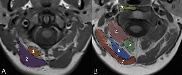
MRI provides detailed anatomical information crucial for understanding various pathologies.
Suboccipital Muscle Anatomy
Brachial Plexus Neurography
The Role of Cervical MRI in Diagnosis and Management
MRI of the cervical spine plays a pivotal role in the diagnosis of a wide spectrum of conditions affecting this region. Its ability to provide detailed images of both osseous and soft tissue structures makes it indispensable for identifying the cause of neck pain, radiculopathy, myelopathy, and other neurological deficits. The information obtained from a cervical MRI is critical for guiding appropriate medical or surgical management, planning interventions, and monitoring treatment outcomes, ultimately contributing to improved patient care and prognosis.
References
- Ross JS, Brant-Zawadzki M, Moore KR, et al. Diagnostic Imaging: Spine. 3rd ed. Amirsys Elsevier; 2015.
- Westbrook C, Roth C, Talbot J. MRI in Practice. 5th ed. Wiley-Blackwell; 2018. Chapter on Spinal Imaging.
- Atlas SW. Magnetic Resonance Imaging of the Brain and Spine. 5th ed. Lippincott Williams & Wilkins; 2016.
- Modic MT, Masaryk TJ, Ross JS, Carter JR. Magnetic resonance imaging of the spine. Radiol Clin North Am. 1986 Mar;24(2):229-45.
- Brant-Zawadzki M, Jensen MC, Obuchowski N, Ross JS, Modic MT. Interobserver variability and diagnostic performance of T2-weighted MR and myelography in the evaluation of cervical radiculopathy. AJNR Am J Neuroradiol. 1995 May;16(5):1057-63.
- American College of Radiology. ACR Appropriateness Criteria® Neck Pain or Cervical Radiculopathy. Last review date: 2022.
- Wilmink JT. CT and MRI of the spine and spinal cord. Saunders Ltd.; 2009.
- Thurnher MM, Law M. Magnetic resonance imaging of the cervical spine. Magn Reson Imaging Clin N Am. 2009 Aug;17(3):357-73.
See also
- Magnetic Resonance Imaging (MRI)
- Magnetic Resonance Angiography (MRA) of the Cerebral Vessels
- Magnetic Resonance Imaging (MRI) of the Abdomen
- Magnetic Resonance Imaging (MRI) of the Brain
- Magnetic Resonance Imaging (MRI) of the Cervical Spine
- Magnetic Resonance Imaging (MRI) of the Hip Joint
- Magnetic Resonance Imaging (MRI) of the Knee Joint
- Magnetic Resonance Imaging (MRI) of the Lumbar Spine
- Magnetic Resonance Imaging (MRI) of the Pelvic Organs
- Magnetic Resonance Imaging (MRI) of the Pituitary Gland (Hypophysis)
- Magnetic Resonance Imaging (MRI) of the Shoulder Joint
- Magnetic Resonance Imaging (MRI) of the Thoracic Cavity Organs
- Magnetic Resonance Imaging (MRI) of the Thoracic Spine
- Magnetic Resonance Imaging (MRI) Study Principle
- Whole-Body Magnetic Resonance Imaging (MRI)


