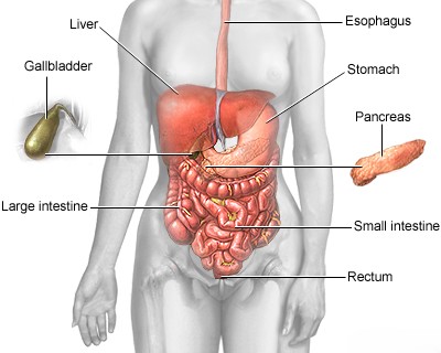
Abdominal CT scan
Abdominal CT scan
Abdominal CT scan should be performed on multislice computed tomography (MSCT) scanners, which allow for rapid acquisition of high-resolution images.
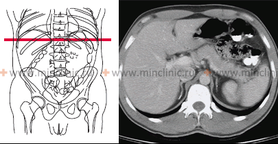
Abdominal CT scan - covering the liver, gallbladder, pancreas system, spleen, kidneys, adrenal glands, stomach, intestines, and major blood vessels - is often carried out after a preliminary ultrasound examination of this anatomical region. It is advisable when undergoing computed tomography (CT scans) of the abdominal cavity to bring along conclusions and images obtained from previous studies, such as ultrasound, even if no pathology was initially found, as CT can provide more detailed information.
Abdominal CT scan (including liver and pancreas) is indicated for:
- Suspected primary or secondary malignant lesions (metastases) of the liver and bile ducts.
- Evaluation of benign liver conditions like fatty liver (steatosis), abscesses, cysts (including parasitic cysts like hydatid disease), and other masses.
- Assessment of liver cirrhosis and its complications (e.g., portal hypertension, hepatocellular carcinoma screening).
- Investigation of obstructive jaundice causes (e.g., stones, strictures, tumors).
- Monitoring the effectiveness of treatment for abdominal tumors.
- Evaluating unexplained hepatomegaly (enlarged liver) or splenomegaly (enlarged spleen).
- Assessing traumatic injuries to abdominal organs (liver, spleen, kidneys, bowel).
- Diagnosing hepatocerebral dystrophy (Wilson's disease).
- Investigating cholelithiasis (gallstones), particularly for suspected stones in the common bile duct (choledocholithiasis).
- Diagnosing acute and chronic pancreatitis and their complications (e.g., pseudocysts, necrosis).
- Suspected neoplasms (tumors) of the pancreas, primary or secondary.
- Evaluating ischemic conditions affecting parenchymal abdominal organs (e.g., bowel ischemia, renal infarction).
- Investigating unexplained abdominal pain, weight loss, or fever.
Abdominal CT scan (liver, gallbladder, pancreas, and related systems) is a crucial method for differentiating between various conditions affecting the parenchymal structures of the abdominal cavity and retroperitoneal space.
Currently, contrast-enhanced abdominal CT scan (liver, gall bladder, pancreas) is considered the most informative non-invasive imaging method for the diagnosis and differential diagnosis of focal liver lesions such as metastases, hemangiomas, adenomas, focal nodular hyperplasia (FNH), hepatocellular carcinoma (HCC), and other tumors or tumor-like processes.
Abdominal CT scan clearly reveals both solid (dense) and cystic tumors of the pancreas, helping to characterize them and assess their extent.
A valuable complement to standard abdominal CT, particularly for biliary issues, is Magnetic Resonance Cholangiopancreatography (MRCP), although CT can also provide information about the bile ducts, especially with contrast. MRCP uses MRI technology to create detailed images of the hepatobiliary and pancreatic ducts without ionizing radiation or invasive procedures. It is a non-invasive alternative to diagnostic Endoscopic Retrograde Cholangiopancreatography (ERCP), which carries a risk of complications like pancreatitis.
CT cholangiography (imaging the bile ducts using CT, usually with contrast) can be used successfully in the diagnosis of anomalies, bile duct strictures, suspected sclerosing cholangitis, and choledocholithiasis (detection of stones in the gallbladder and bile ducts), although MRCP is often preferred for non-calcified stones and ductal evaluation.
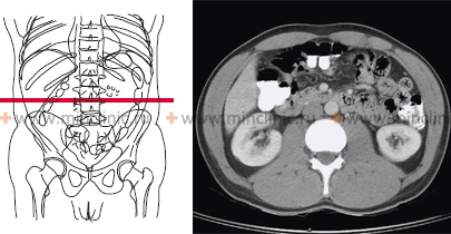
Indications for computed tomography (CT) scan specifically focused on the kidneys and adrenal glands include:
- Patients with contraindications to intravenous contrast for excretory urography (e.g., severe allergy to iodinated contrast media, severe renal impairment).
- To clarify the nature of renal or adrenal masses detected by other methods (e.g., ultrasound), differentiating cysts from solid tumors or normal variants.
- Patients with clinical findings suggestive of renal tumors (e.g., hematuria, flank pain, palpable mass).
- For diagnosis and staging of renal cell carcinoma and other kidney tumors.
- Evaluation of suspected adrenal tumors (adenoma, pheochromocytoma, carcinoma, metastasis).
- Diagnosis of pathological processes surrounding the kidney (paranephric space), such as abscesses or hematomas.
- Assessment of suspected congenital abnormalities of the urinary system.
- Evaluation for kidney stones (urolithiasis), especially non-contrast CT ("CT KUB").
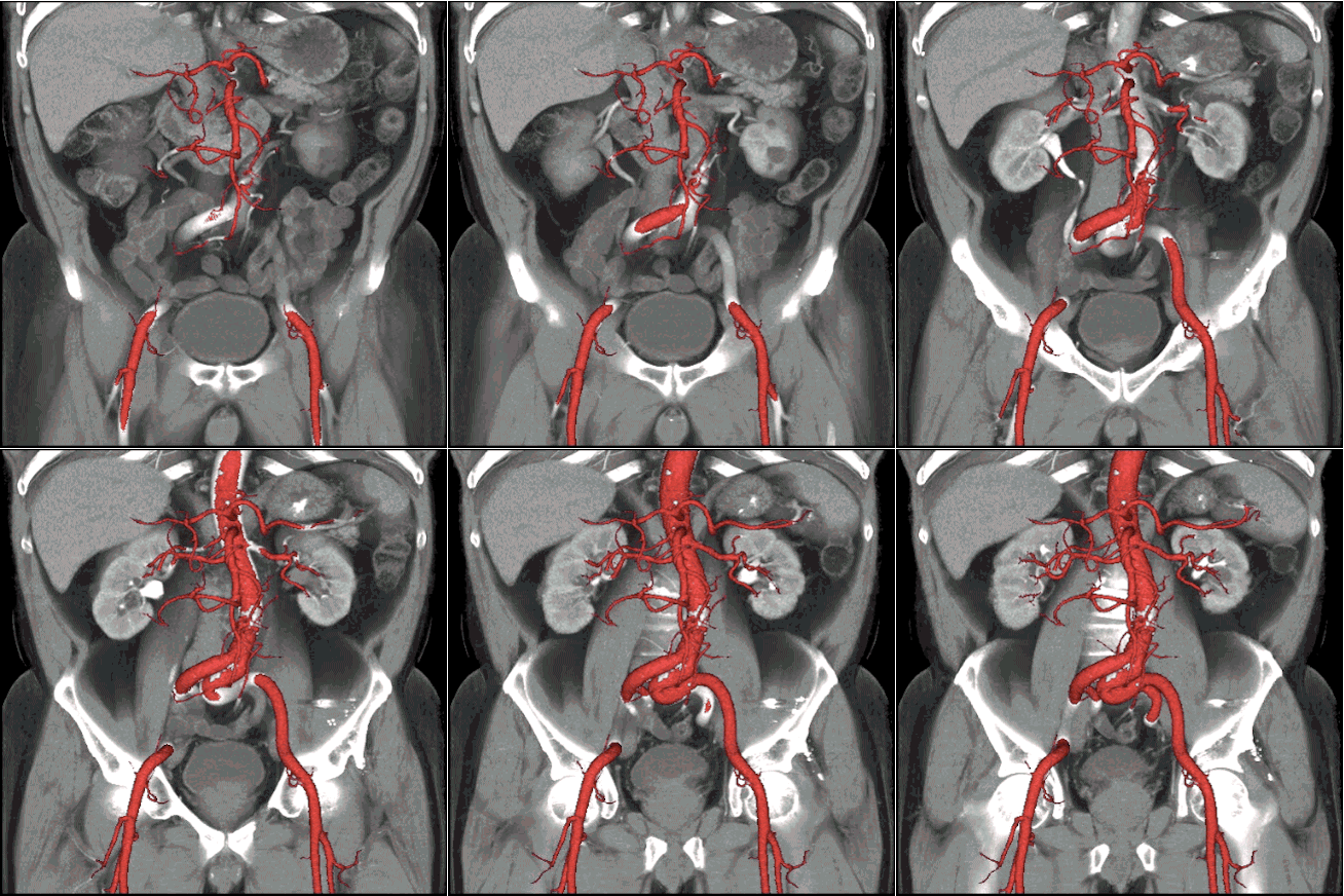
- Assessment of renal vascular disease (using CT angiography).
Computed tomography (CT) scan of the kidneys and adrenal glands is a highly effective diagnostic method. It allows for high accuracy in differentiating adrenal masses (malignant vs. benign) using specific imaging protocols, including assessing density on non-contrast scans and measuring contrast washout, which are sensitive indicators for the presence of intracellular fat typical in benign adenomas.
Preparation for the Scan
Preparation for an abdominal CT scan can vary depending on the reason for the scan and whether contrast material will be used.
- Fasting: You may be asked not to eat or drink for several hours before the scan, especially if intravenous (IV) contrast is planned. This reduces the risk of aspiration if nausea occurs from the contrast.
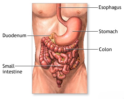
- Oral Contrast: For some scans evaluating the stomach or bowels, you might need to drink a special contrast liquid starting 1-2 hours before the procedure. This helps outline the digestive tract on the images. Water may sometimes be used as a negative contrast agent.
- Clothing: Wear comfortable, loose-fitting clothing. You may be asked to change into a hospital gown. Metal objects like jewelry, eyeglasses, dentures, and hairpins can interfere with the images and should be removed.
- Medications: Inform your doctor about all medications you are taking and if you have any allergies, especially to iodine or previous contrast reactions. Specific instructions may be given for certain medications, like metformin (used for diabetes), if IV contrast is used.
- Medical Conditions: Notify your doctor and the CT technologist if you have any serious medical conditions, such as heart disease, asthma, diabetes, kidney disease, or thyroid problems. It's also crucial to inform them if there's any possibility you might be pregnant.
The CT Procedure
During the CT scan:
- You will lie on a narrow table that slides into the center of the large, doughnut-shaped CT scanner.
- Pillows and straps may be used to help you stay still and maintain the correct position during the scan, as movement can blur the images.
- If IV contrast is needed, a small IV line will be placed in a vein in your arm or hand before the scan begins.
- The technologist will operate the scanner from an adjacent room but can see, hear, and speak with you through an intercom system.
- As the scanner works, the table will move slowly through the machine. You might hear whirring or buzzing sounds.
- You may be asked to hold your breath for short periods (usually 10-20 seconds) during image acquisition to minimize motion artifacts, especially from breathing.
- The entire procedure is usually painless and typically takes about 10 to 30 minutes, although preparation time might add to the overall duration.
Use of Contrast Material
Contrast materials, often iodine-based for CT, are substances used to improve the visibility of internal body structures. They can be administered in three ways for abdominal CT:
- Oral Contrast: Swallowed liquid used to highlight the esophagus, stomach, and intestines.
- Rectal Contrast: Administered via an enema to visualize the large intestine or rectum.
- Intravenous (IV) Contrast: Injected into a vein to enhance blood vessels, kidneys, liver, spleen, and other organs, helping to differentiate normal from abnormal tissues (like tumors or inflammation). When IV contrast is injected, you might feel a warm sensation throughout your body and possibly a metallic taste in your mouth; these effects are usually temporary and harmless.
The decision to use contrast depends on the clinical question being asked.
Risks and Benefits
Benefits:
- CT scans are fast, painless, non-invasive, and accurate.
- They provide highly detailed images of many types of tissue (bone, soft tissue, blood vessels) simultaneously.
- CT is less sensitive to patient movement than MRI.
- It can be performed even if a patient has implanted medical devices of any kind, unlike MRI.
- CT provides real-time imaging, making it useful for guiding procedures like biopsies and fluid drainages.
Risks:
- Radiation Exposure: CT scans use ionizing radiation (X-rays). While the dose for a single scan is generally low, repeated scans accumulate radiation exposure over time, which carries a small theoretical risk of increasing cancer potential later in life. The benefit of an accurate diagnosis generally outweighs this risk, especially in adults. Doses are kept as low as reasonably achievable (ALARA principle).
- Contrast Reaction: Allergic-like reactions to iodine-based IV contrast material can occur, ranging from mild (itching, hives) to severe (anaphylaxis), although severe reactions are rare. Patients with previous reactions or significant allergies are at higher risk. Kidney problems can sometimes be worsened by IV contrast, especially in patients with pre-existing renal impairment. Proper screening helps minimize this risk.
- Pregnancy: CT scans are generally not recommended for pregnant women unless medically necessary due to potential radiation risk to the fetus. Alternative imaging like ultrasound or MRI might be considered.
References
- Radiological Society of North America (RSNA) & American College of Radiology (ACR). (2023). Body CT (Abdomen and Pelvis). RadiologyInfo.org. Retrieved from https://www.radiologyinfo.org/en/info/bodyct
- Mayo Clinic Staff. (2022). CT scan. Mayo Clinic Patient Care & Health Information. Retrieved from https://www.mayoclinic.org/tests-procedures/ct-scan/about/pac-20393675
- Sahani, D. V., & Samir, A. E. (2017). Abdominal Imaging (2nd ed.). Elsevier. [Note: This is a textbook reference, representing comprehensive information available in medical literature.]
- American College of Radiology. (2023). ACR Manual on Contrast Media. Retrieved from https://www.acr.org/Clinical-Resources/Contrast-Manual
- National Institute of Biomedical Imaging and Bioengineering (NIBIB). (n.d.). Computed Tomography (CT). NIH - National Institutes of Health. Retrieved from https://www.nibib.nih.gov/science-education/science-topics/computed-tomography-ct

