Anatomy of the nervous system
Introduction to the Nervous System
The nervous system is the body's primary command and communication network, responsible for coordinating all voluntary and involuntary actions, processing sensory information, and enabling higher cognitive functions such as thought, memory, and emotion. Its intricate anatomy allows for rapid transmission of electrochemical signals throughout the body.
Major Divisions: CNS and PNS
The nervous system is broadly divided into two main parts:
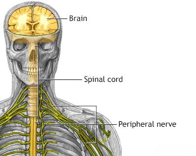
- The Central Nervous System (CNS): This is the control center, consisting of the brain and the spinal cord. It processes information and coordinates bodily functions.
- The Peripheral Nervous System (PNS): This comprises all the nervous tissue outside the CNS, including cranial nerves (originating from the brain), spinal nerves (originating from the spinal cord), and their associated ganglia. The PNS connects the CNS to the limbs and organs, essentially serving as the communication relay going back and forth between the brain and the extremities.
The Central Nervous System (CNS)
The Brain (Encephalon)
The brain, housed within the protective cranial cavity, is the most complex part of the nervous system. It is responsible for interpreting senses, initiating body movement, and controlling behavior. Major components include:
Cerebrum: Lobes and Functional Areas
The cerebrum is the largest part of the brain, divided into two hemispheres (left and right), which are connected by the corpus callosum. Its highly convoluted surface, the cerebral cortex, is responsible for higher-level cognitive functions.
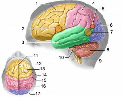
Functional anatomy of the human brain, illustrating key structures: 1 – frontal lobe, 2 – motor zone of Broca's speech center, 3 – temporal lobe, 4 – parietal lobe, 5 – reading perception zone, 6 – occipital lobe, 7 – sensory zone of Wernicke's speech center, 8 – cerebellum, 9 – medulla oblongata, 10 – pons, 11 – longitudinal fissure, 12 – (another view of) frontal lobe, 13 – premotor zone, 14 – precentral gyrus (primary motor cortex), 15 – postcentral gyrus (primary somatosensory cortex), 16 – (another view of) parietal lobe, 17 – (another view of) occipital lobe.
Each hemisphere is further divided into lobes, each associated with specific functions:
- Frontal Lobe (1, 12): Located at the front of the brain, it's involved in planning, decision-making, working memory, voluntary movement (motor cortex/precentral gyrus - 14), personality, and expressive language (Broca's area - 2, typically in the left hemisphere). The premotor zone (13) is involved in planning movements.
- Parietal Lobe (4, 16): Positioned behind the frontal lobe, it processes sensory information such as touch, temperature, pain, and pressure (somatosensory cortex/postcentral gyrus - 15). It's also involved in spatial awareness, navigation, and attention. The reading perception zone (5) is often located here.
- Temporal Lobe (3): Located beneath the lateral fissure, it's crucial for processing auditory information, memory formation, language comprehension (Wernicke's area - 7, typically in the left hemisphere), and object recognition.
- Occipital Lobe (6): Situated at the back of the brain, it is the primary visual processing center.
The longitudinal fissure (11) is the deep groove that separates the two cerebral hemispheres.
Cerebellum (8)
Located at the back and base of the brain, beneath the occipital and temporal lobes, the cerebellum is crucial for coordinating voluntary movements, posture, balance, motor learning, and some cognitive functions.
Brainstem (Medulla Oblongata, Pons)
The brainstem connects the cerebrum and cerebellum to the spinal cord. It controls essential life-sustaining functions:
- Medulla Oblongata (9): The lowest part of the brainstem, controlling autonomic functions like breathing, heart rate, and blood pressure.
- Pons (10): Located above the medulla, it relays signals from the forebrain to the cerebellum and is involved in sleep, respiration, swallowing, bladder control, hearing, equilibrium, taste, eye movement, facial expressions, and sensation.
- Midbrain (not explicitly numbered in image caption but superior to pons): Involved in vision, hearing, motor control, sleep/wake, arousal (alertness), and temperature regulation.
The Spinal Cord
The spinal cord is a long, cylindrical structure extending from the brainstem down the vertebral canal. It transmits nerve signals between the brain and the rest of the body and controls reflexes. It is protected by the vertebral column and surrounded by cerebrospinal fluid and meninges.
The Peripheral Nervous System (PNS)
The PNS includes all neural structures outside the brain and spinal cord. Its main role is to connect the CNS to the limbs, organs, and skin.
Cranial Nerves
There are 12 pairs of cranial nerves that emerge directly from the brain and brainstem. They are responsible for sensory and motor functions primarily in the head and neck region (e.g., vision, smell, hearing, facial movement, taste).
Spinal Nerves and Segmental Innervation
There are 31 pairs of spinal nerves that emerge from the spinal cord and are named according to the vertebral region from which they exit (cervical, thoracic, lumbar, sacral, coccygeal). Each spinal nerve provides sensory and motor innervation to a specific segment of the body (dermatome for sensory, myotome for motor).
Diagram illustrating the segmental innervation pattern of the human body by spinal nerves. Different colors represent nerves originating from distinct spinal regions: green often indicates cervical nerves, blue for thoracic nerves, lilac for lumbar nerves, and pink for sacral nerves, mapping to dermatomes and myotomes.
The Autonomic Nervous System (ANS)
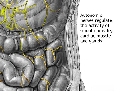
The ANS is a division of the PNS that controls involuntary bodily functions such as heart rate, digestion, respiratory rate, pupillary response, urination, and sexual arousal. It is further divided into:
- Sympathetic Nervous System: Generally responsible for the "fight or flight" response.
- Parasympathetic Nervous System: Generally responsible for the "rest and digest" response.
The ANS innervates internal organs like the stomach and intestines, regulating their activity without conscious control.
Cellular Components: Neurons and Glia
The nervous system is composed of two main types of cells:
- Neurons (Nerve Cells): The fundamental units responsible for transmitting nerve impulses. They consist of a cell body (soma), dendrites (receiving signals), and an axon (transmitting signals).
- Glial Cells (Neuroglia): Supporting cells that provide structural support, insulation (myelin), nourishment, and immune defense for neurons. Types include astrocytes, oligodendrocytes, microglia, and Schwann cells (in the PNS).
Key Functional Systems
The nervous system operates through complex interconnected pathways forming various functional systems.
Sensory Pathways
These pathways transmit information from sensory receptors throughout the body (e.g., for touch, pain, temperature, vision, hearing, smell, taste) to the CNS for processing.
Motor Pathways
These pathways carry signals from the CNS to muscles and glands, initiating voluntary movements (somatic motor system) and controlling involuntary functions (autonomic motor system).
Speech and Language Centers
Specialized areas in the brain, primarily in the left hemisphere for most individuals, are dedicated to language processing. Broca's area (typically in the frontal lobe) is involved in speech production, while Wernicke's area (typically in the temporal lobe) is crucial for language comprehension.
Visual Pathway
This involves the eyes, optic nerves (Cranial Nerve II), optic chiasm, optic tracts, lateral geniculate nuclei of the thalamus, and the visual cortex in the occipital lobes, processing visual information.
Comparative Overview: CNS vs. PNS
| Feature | Central Nervous System (CNS) | Peripheral Nervous System (PNS) |
|---|---|---|
| Components | Brain and Spinal Cord | Cranial nerves, Spinal nerves, Ganglia, Autonomic nervous system components outside CNS |
| Primary Function | Integration and command center; processes information, coordinates activity | Transmits sensory information to CNS and motor commands from CNS to effectors (muscles, glands) |
| Protection | Protected by bone (skull, vertebral column), meninges, and cerebrospinal fluid | Less protected; more vulnerable to trauma and toxins |
| Regeneration Capacity | Limited regeneration of damaged neurons | Greater capacity for axon regeneration (if cell body is intact and Schwann cells are present) |
| Supporting Cells (Glia) | Astrocytes, Oligodendrocytes, Microglia, Ependymal cells | Schwann cells (form myelin), Satellite cells (support neuron cell bodies in ganglia) |
Clinical Relevance and Related Pathologies (Index)
Understanding the anatomy of the nervous system is crucial for diagnosing and treating a wide range of neurological disorders. The collapsible sections below provide links to articles on specific diseases affecting the brain and peripheral nerves.
Brain Diseases Index
This section serves as an index to related articles on specific brain diseases.
- Central nervous system infections:
- Brain abscess (lobar, cerebellar)
- Eosinophilic granuloma, Langerhans cell histiocytosis (LCH), Hennebert's symptom
- Epidural brain abscess
- Sinusitis-associated intracranial complications
- Otogenic intracranial complications
- Sinusitis-associated ophthalmic complications
- Bacterial otogenic meningitis
- Subdural brain abscess
- Sigmoid sinus suppurative thrombophlebitis
- Cerebral 3rd Ventricle Colloid Cyst
- Cerebral and spinal adhesive arachnoiditis
- Corticobasal Ganglionic Degeneration (Limited Brain Atrophy)
- Encephalopathy
- Headache, migraine
- Traumatic brain injury (concussion, contusion, brain hemorrhage, axonal shearing lesions)
- Increased intracranial pressure and hydrocephalus
- Parkinson's disease
- Pituitary microadenoma, macroadenoma and nonfunctioning adenomas (NFPAs), hyperprolactinemia syndrome
- Spontaneous cranial cerebrospinal fluid leak (CSF liquorrhea)
Peripheral Nerve Diseases Index
This section serves as an index to related articles on specific peripheral nerve diseases.
- Spinal disc herniation (often causing peripheral nerve root compression)
- Pain in the arm and neck (trauma, cervical radiculopathy)
- The eyeball and the visual pathway (involving cranial nerves):
- Optic nerve (Cranial Nerve II) and retina:
- Compression neuropathy of the optic nerve
- Edema of the optic disc (papilledema)
- Ischemic neuropathy of the optic nerve
- Meningioma of the optic nerve sheath
- Optic nerve atrophy
- Optic neuritis in adults
- Optic neuritis in children
- Opto-chiasmal arachnoiditis (affecting optic nerves/chiasm)
- Pseudo-edema of the optic disc (pseudopapilledema)
- Toxic and nutritional optic neuropathy
- Neuropathies and neuralgia:
- Diabetic, alcoholic, toxic and small fiber sensory neuropathy (SFSN)
- Facial nerve (Cranial Nerve VII) neuritis (Bell's palsy, post-traumatic neuropathy)
- Fibular (peroneal) nerve neuropathy
- Median nerve neuropathy
- Neuralgia (intercostal, occipital, facial, glossopharyngeal, trigeminal, metatarsal)
- Radial nerve neuropathy
- Tibial nerve neuropathy
- Traumatic (post-traumatic) neuropathies
- Traumatic (post-traumatic) trigeminal (Cranial Nerve V) neuropathy
- Traumatic neuropathy of the sciatic nerve
- Trigeminal neuralgia
- Ulnar nerve neuropathy
- Tumors (neoplasms) of the peripheral nerves and autonomic nervous system (neuroma, sarcomatosis, melanoma, neurofibromatosis, Recklinghausen's disease)
- Carpal tunnel syndrome (Median nerve compression)
- Ulnar nerve compression in the cubital canal (Cubital tunnel syndrome)
References
- Kandel ER, Schwartz JH, Jessell TM, Siegelbaum SA, Hudspeth AJ, Mack S. Principles of Neural Science. 6th ed. McGraw-Hill Education; 2021.
- Brodal P. The Central Nervous System: Structure and Function. 5th ed. Oxford University Press; 2016.
- Nolte J. The Human Brain: An Introduction to its Functional Anatomy. 7th ed. Mosby; 2014.
- Purves D, Augustine GJ, Fitzpatrick D, et al., eds. Neuroscience. 6th ed. Sinauer Associates; 2018.
- Snell RS. Clinical Neuroanatomy. 8th ed. Lippincott Williams & Wilkins; 2019.
- Blumenfeld H. Neuroanatomy through Clinical Cases. 3rd ed. Sinauer Associates; 2021.
- Gray H. Gray's Anatomy: The Anatomical Basis of Clinical Practice. 42nd ed. Elsevier; 2020. (Relevant sections on neuroanatomy).
See also
- Glossary - provides a list of the most common medical terms
- A dictionary of neurological signs - neurological signs and their meaning;
- Frequently Asked Questions - lists the most frequently asked questions by patients
- Anatomy of the spine - basic data on the structure of the spine and spinal cord are given and illustrated
- Anatomy of the nervous system - basic data on the structure of the brain and spinal cord, nerves are given and illustrated
- Rules for preparing the person under study for certain diagnostic procedures
- Nursing rules - presents ways to safely care for bedridden or debilitated patients at home or in a hospital


