Endoscopic Treatment of Ulnar Nerve Compression in the Cubital Canal
Endoscopic treatment of ulnar nerve compression in the cubital canal
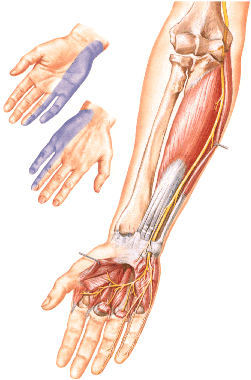
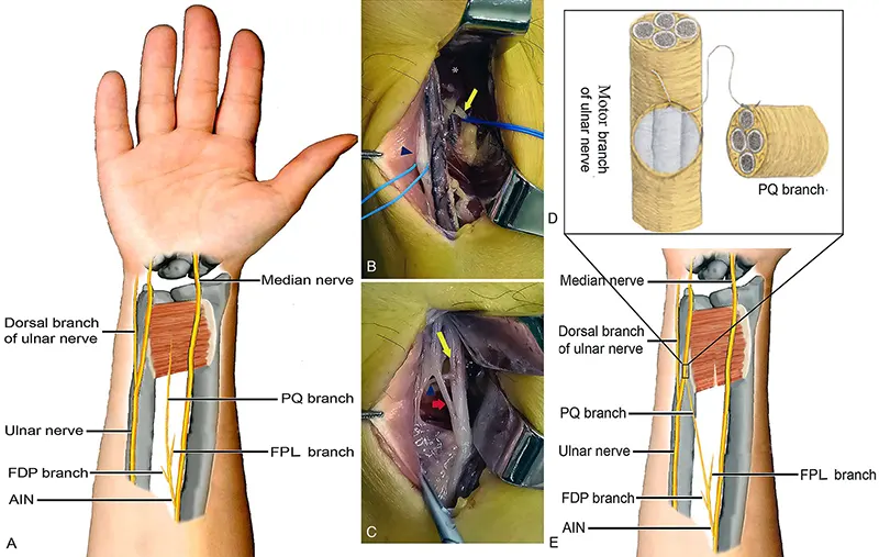
Syndrome of the cubital canal (Sulcus Ulnaris Syndrome) is a compression of the nerve of the upper limb and is the second most common after carpal tunnel syndrome (carpal tunnel).
With traditional methods, a large skin incision of 8 to 12 cm is required, and in many cases, the nerve is still moved to an inappropriate physiological position.
Thanks to the endoscopic method, it is possible to reduce not only the skin incision to 1.5–2 cm, but also significantly increase the area of neurolysis (15–20 cm). In this case, it is possible to divide all fibrous arcades between the heads of M. flexor carpi ulnaris and thus achieve a much more complete decompression of the nerve than with the old methods.
In the endoscopic method, a subcutaneous space is created using the forceps used as tunnel forceps. In this case, first, a mirror-illuminator is inserted, then a 4 mm endoscope with a dissector at the end. Preparation is carried out under optical control.
The advantages of endoscopic surgery in comparison with previous methods are as follows:
- low incidence (in over 90% of patients, elbow mobility is restored within 24 hours)
- rapid improvement of symptoms (in over 95% of patients within 24 hours)
- fast recovery
Our experience, as a result of over 100 interventions, shows that this method is reliable, easy to learn, and an excellent alternative to the surgical methods that existed until now.
The instrumentation has also proven itself suitable for endoscopic and minimally invasive operations for Epicondylitis humeri radailis and ulnaris, compression of the radial nerve on the forearm (Wartenberg syndrome), and pronator syndrome.


