Arthroscopy of the ankle joint
- Arthroscopy of the ankle joint
- Anatomy of the ankle joint
- Indications for arthroscopy of the ankle joint
- arthrosis of the ankle joint
- arthrofibrosis of the ankle joint
- posterior impingement of the ankle joint
- instability of the ankle joint
- osteochondral defects of the ankle joint
- anterior ankle impingement
- synovitis of the ankle joint
- condition after ankle fractures
- articular (chondromic) bodies in the ankle joint
- The advantages of arthroscopy of the ankle joint
Arthroscopy of the ankle joint
Arthroscopy of the ankle joint is a surgical procedure in which both diagnostics and therapeutic manipulation are performed in the event of damage to the structures of the ankle joint. During arthroscopy of the ankle joint, 2 small, thinnest skin approaches are performed, through which, with the help of optics and special arthroscopic instruments, a therapeutic procedure is performed inside the cavity of the ankle joint.
Arthroscopy of the ankle joint is a minimally invasive surgical procedure used in orthopedics to treat problems in the ankle joint. Arthroscopy of the ankle joint is performed using a thin camera (arthroscope) and fiber optic illumination. The arthroscope can magnify and transmit the video image taken inside the joint cavity to a monitor.
The operation aims to reduce pain symptoms and improve the function of the ankle joint in the patient.
Anatomy of the ankle joint
The ankle joint is formed by the connection of three bones. The talus is placed in the so-called fork, which is formed by the fibula and tibia from above. Under the talus is the calcaneus, together they form the subtalar joint. Ligaments are dense strands that hold bones together. The lateral ligament complex outside the ankle joint is represented by three ligaments: the anterior talofibular ligament, the calcaneofibular ligament, and the posterior talofibular ligament.
Ankle ligament rupture or inversion trauma (internal dislocation) of the ankle joint most often involves damage to the anterior talofibular and calcaneofibular ligaments.
The implant is placed in the intervertebral space in a folded state (in the form of a rod with a diameter of 5 mm) using a special device. It also allows, after installation, to push out of the rod metal segments - teeth. Two implants are placed in one intervertebral space.
The anterior talofibular ligament performs several functions:
- keeps the ankle and talus from displacement anteriorly
- provides both anteroposterior and lateral stability of the ankle
- limits flexion and inversion of the foot
- prevents internal rotation of the talus
The calcaneofibular ligament has the following functions:
- keeps the ankle joint from shifting inwards
- limits inversion of the foot
- prevents excessive extension of the foot
- prevents internal rotation of the talus bone
The posterior talofibular ligament is the strongest of the three external lateral ligaments. This ligament is almost entirely surrounded by the synovial membrane. Functions of the posterior talofibular ligament:
- prevents the extension of the foot
- restricts the rear offset
- restricts the external rotation of the talus bone
- the short fibers of the ligament restrain excessive adduction of the foot
On the inner side, in the form of a triangular shape, there is a deltoid ligament. The deltoid ligament plays a major role in stabilizing the ankle joint. The deltoid ligament performs the following functions:
- restrains excessive eversion (turning outwards) movements of the foot
- restrains flexion, pronation and external rotation of the foot
- prevents the hallux valgus (external) inclination of the talus bone
Indications for arthroscopy of the ankle joint
Arthroscopy can be used to diagnose and treat various diseases of the ankle joint. Diseases that can be treated with this technology include:
- arthrosis of the ankle joint
- arthrofibrosis of the ankle joint
- dislocation of the foot
- posterior impingement of the ankle joint
- instability of the ankle joint
- osteochondral defects of the ankle joint
- acute and chronic ankle injury
- anterior ankle impingement
- damage to the ligaments of the ankle joint
- synovitis of the ankle joint
- condition after ankle fractures
- sports injuries of the ankle joint
- articular (chondromic) bodies in the ankle joint
Arthrosis of the ankle joint
This treatment option is suitable for many patients as a minimally invasive method, is more effective and alternative than the open method of surgical treatment.
Arthrofibrosis
Scar tissue can form in the ankle joint. This can lead to soreness and limited range of motion in the ankle joint. Arthroscopy of the ankle joint can be used to identify scar tissue and remove it (excision).
Posterior impingement of the ankle joint
Posterior impingement occurs when the soft tissues in the back of the ankle joint become inflamed and subsequently thicken. Patients may feel soreness with plantar flexion of the foot. Most often, this syndrome usually occurs in those engaged in dancing, ballet, with prolonged walking in high heels. It may also be related to:
- pronounced posterolateral process (Stida process)
- as a result of acute trauma, fracture of the posterior process of the talus
- avulsion (detachment) of the posterior talofibular ligament
Damaged and altered tissues can be removed by arthroscopy.
Instability of the ankle joint
The ligaments of the ankle joint are often damaged as a result of injuries. This can lead to a feeling of instability, twisting of the foot when walking, especially on uneven surfaces, the appearance of pain, swelling in the ankle joint.
The ligaments of the ankle joint can be restored with the help of arthroscopic surgical techniques. Arthroscopic techniques can be an option for this task, which will help to stabilize the ankle joint.
Osteochondral defects
These are areas of damaged cartilage and bone in the area of the ankle joint, which are also caused by trauma (fractures and dislocations). Common symptoms include pain and swelling of the ankle joint. Patients may complain of pain in the extreme positions of the foot and clicks in the ankle joint. Diagnosis of an osteochondral defect includes clinical methods of investigation, visualization may include X-rays,MRI or CT. Treatment depends on the size and location of the osteochondral defect. Arthroscopic surgery allows you to remove damaged cartilage, and perform perforation (drilling small holes in the bone) to accelerate healing. Cartilage transplantation is also performed.
Anterior ankle impingement (footballer's ankle joint)
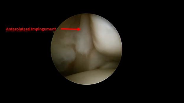
Impingement occurs when the bone or soft tissue on the front of the ankle joint becomes inflamed. Symptoms include pain and swelling of the ankle joint. This may limit the back flexion in the ankle joint. Patients often experience pain when walking uphill. Osteophytes (bone growths) in the anterior part of the ankle joint can be seen on an X-ray. Arthroscopy can be used to remove inflamed tissues and bone growths (osteophytes).
Synovitis
Synovitis is an inflammation of the mucous membrane of the ankle joint that causes pain and swelling. Synovitis can be caused by trauma. In inflammatory arthritis (rheumatoid arthritis) arthroscopy of the ankle joint can be used to surgically remove significantly enlarged inflammatory tissue that is no longer amenable to conservative treatment.
Condition after ankle fractures
Arthroscopy of the ankle joint can be used in the long-term period in patients after surgical treatment of the ankles. After an ankle fracture, in 85% of cases, the cartilage covering the bone in the joint is damaged. When the cartilage surface is damaged, there is an impaired synthesis of synovial fluid inside the joint cavity, the cartilage is detached into the joint cavity, pain, swelling, and restriction of movement in the ankle joint occur.
Articular chondromic bodies
Articular bodies are formed after injuries in the form of cartilage, bone, and scar tissue, which break away from the place of attachment and become free floating bodies in the joint cavity. Joint bodies can cause pain when the foot is positioned. Arthroscopy of the ankle joint in this case will help eliminate the chondromic bodies and provide free movement in the joint.
The advantages of arthroscopy of the ankle joint
Hospital stay after arthroscopy of the ankle joint is from 3 to 7 days, depending on the nature of the damage to the ankle joint.
Arthroscopy of the ankle joint allows patients to start rehabilitation and return to high-level activity activities, such as sports or heavy physical labor, faster than with open surgeries. With arthroscopy, there are fewer cosmetic postoperative defects.
Full recovery of the ankle joint function depends on the type of surgery. Rehabilitation includes specially developed physiotherapy procedures and a specially designed set of physical therapy exercises that will help control pain and swelling after surgery, as well as improve the range and strength of movement in the ankle joint.
Rehabilitation arthroscopy of the ankle joint
Sometimes patients develop symptoms that cannot be investigated by other diagnostic methods. Arthroscopy makes it possible to examine the condition of the ankle joint in detail and identify the cause of the existing symptoms.
Results of ankle arthroscopy: 70 to 90% of patients who underwent ankle arthroscopy with the most common joint problems received good or excellent treatment results.
See also
- Achilles tendon inflammation (paratenonitis, ahillobursitis)
- Achilles tendon injury (sprain, rupture)
- Ankle and foot sprain
- Arthritis and arthrosis (osteoarthritis):
- Autoimmune connective tissue disease:
- Bunion (hallux valgus)
- Epicondylitis ("tennis elbow")
- Hygroma
- Joint ankylosis
- Joint contractures
- Joint dislocation:
- Knee joint (ligaments and meniscus) injury
- Metabolic bone disease:
- Myositis, fibromyalgia (muscle pain)
- Plantar fasciitis (heel spurs)
- Tenosynovitis, infectious tenosynovitis
- Vitamin D and parathyroid hormone


