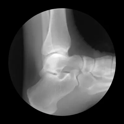
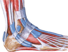
Foot and ankle dislocation
Foot & Ankle Dislocation Overview
Dislocations involving the foot and ankle encompass several distinct injury patterns [1, 2]:
- Ankle (Tibiotalar) Joint Dislocation: Displacement of the talus bone relative to the tibia and fibula.
- Isolated Talus Dislocation: The talus bone displaces from both the ankle joint above and the subtalar joint below.
- Subtalar Dislocation: Dislocation occurs at the joint between the talus and the calcaneus (heel bone) and the talus and the navicular bone, while the ankle joint remains intact. The rest of the foot displaces relative to the talus.
- Midfoot Dislocations: Involving the Chopart joint (talonavicular and calcaneocuboid) or Lisfranc joint (tarsometatarsal).
- Toe Dislocations: Affecting the metatarsophalangeal (MTP) or interphalangeal (IP) joints.
Ankle (Tibiotalar) Joint Dislocation: Pure dislocations of the ankle joint without associated fractures are rare because the mortise (the socket formed by the tibia and fibula) provides strong bony stability [1, 2]. Most ankle dislocations occur in conjunction with malleolar fractures (fractures of the distal tibia and/or fibula), such as complex ankle fracture-dislocations (e.g., Dupuytren fracture) [1, 2].
- Posterior Ankle Dislocation (Most Common): Usually results from severe plantar flexion force, often with associated malleolar fractures [1]. The foot appears shortened, and the heel prominence is reduced [1].
- Anterior Ankle Dislocation: Results from forced dorsiflexion, often associated with anterior tibial rim fractures [1]. The hindfoot appears elongated [1].
- Lateral/Medial Ankle Dislocation: Require significant force and are almost always associated with severe malleolar fractures [1].
Lateral Foot Dislocation & Ankle Fractures
Significant lateral (sideways) displacement of the foot at the ankle level almost invariably involves fractures of the malleoli (the bony prominences on either side of the ankle) [1, 2]. A pure lateral ankle dislocation without fracture is practically impossible due to the strong bony constraints of the ankle mortise and ligamentous support [1].
Injuries like the Dupuytren fracture (a specific type of ankle fracture-dislocation involving fibular fracture, syndesmotic ligament disruption, and often medial malleolus fracture or deltoid ligament rupture) are commonly associated with lateral displacement or instability of the talus within the mortise [1, 2]. The foot typically assumes a position shifted laterally (valgus) or medially (varus) relative to the leg [1].
Reduction of ankle dislocations, especially those without severe fractures ("pure" or simple dislocations, though rare), can sometimes be achieved relatively easily with traction and manipulation, particularly under anesthesia to overcome muscle spasm [1]. However, the presence of associated fractures often necessitates open reduction and internal fixation (ORIF) to restore joint congruity and stability [1, 2].
Talus (Talar Bone) & Subtalar Dislocation
Isolated Talus Dislocation: A rare but severe injury where the talus bone is completely extruded from its articulations with the tibia/fibula (ankle joint), calcaneus (subtalar joint), and navicular (talonavicular joint) [1, 3]. This requires rupture of multiple strong ligaments and significant trauma (e.g., high-energy falls, motor vehicle accidents) [1]. The talus can displace in various directions (anterior, posterior, medial, lateral) and may even rotate [1]. Associated fractures, particularly of the talar neck, are common [1]. Reduction can be difficult due to the degree of displacement and soft tissue disruption [1]. Open reduction is often necessary, and there's a very high risk of avascular necrosis (AVN) of the talus due to its precarious blood supply [1, 3]. Talar extrusion (open dislocation) is also possible [1].
Subtalar Dislocation: This involves simultaneous dislocation at the talocalcaneal and talonavicular joints, while the tibiotalar (ankle) joint remains intact [1, 3]. The calcaneus and navicular (and the rest of the foot) displace together relative to the talus [1]. It typically results from high-energy trauma involving forceful inversion or eversion of the foot [1, 3].
- Medial Subtalar Dislocation (Most Common): Occurs with forceful inversion, causing the foot to displace medially relative to the talus ("acquired clubfoot" deformity) [1, 3].
- Lateral Subtalar Dislocation: Occurs with forceful eversion, causing the foot to displace laterally ("acquired flatfoot" deformity) [1, 3].
- Anterior/Posterior Subtalar Dislocations: Extremely rare and usually associated with fractures [1].
Clinically, subtalar dislocations present with gross deformity of the foot, severe pain, and swelling [1]. Ankle joint motion (dorsiflexion/plantarflexion) may be preserved to some extent if tested gently, distinguishing it from an ankle dislocation [1]. Closed reduction under anesthesia is usually successful by applying traction and reversing the mechanism of injury (e.g., eversion pressure for a medial dislocation) [1, 3]. Immobilization in a cast for 4-6 weeks is typical, followed by rehabilitation [1]. The prognosis is generally better than for isolated talar dislocations, but post-traumatic arthritis can still occur [1, 3].
Video potentially illustrating reduction techniques for foot or ankle dislocations, emphasizing traction and manipulation under anesthesia [3].
Rare Types of Foot Bones Dislocations
Other dislocations within the foot are less common but significant [1]:
- Midtarsal (Chopart) Joint Dislocation: Dislocation at the articulation between the hindfoot (talus, calcaneus) and the midfoot (navicular, cuboid). These are rare, high-energy injuries often associated with fractures.
- Tarsometatarsal (Lisfranc) Joint Dislocation: Disruption of the joint between the midfoot (cuneiforms, cuboid) and the bases of the metatarsals. This can range from subtle sprains to complete dislocations, often caused by axial load on a plantarflexed foot or crush injuries. Lisfranc injuries are often missed initially but can lead to significant long-term pain and arthritis if not treated appropriately (often requiring surgical fixation). Diagnosis requires careful examination and weight-bearing X-rays or CT scans.
- Isolated Cuneiform, Navicular, or Cuboid Dislocations: Very rare, usually resulting from severe crush injuries.
- Metatarsophalangeal (MTP) & Interphalangeal (IP) Joint Dislocations: Dislocations of the toe joints occur similarly to finger dislocations, often from stubbing injuries or hyperextension/hyperflexion forces. Reduction is usually straightforward with traction and manipulation.
Diagnosis of midfoot and forefoot dislocations often requires X-rays, and CT scans may be needed for detailed assessment, especially for Lisfranc injuries [1]. Reduction involves traction and manipulation [1]. Immobilization in a cast is typically required for 4-6 weeks, sometimes longer, potentially followed by supportive footwear or orthotics [1].
Complications of foot dislocations can include associated fractures, nerve damage (neuromas), soft tissue injury, chronic pain, instability, and post-traumatic arthritis [1]. Early diagnosis and appropriate management are crucial [1]. Custom orthotics or insoles may be helpful for managing residual pain or alignment issues after healing.
Differential Diagnosis of Acute Foot/Ankle Injury
| Condition | Key Features / Distinguishing Points | Typical Investigations / Findings |
|---|---|---|
| Ankle (Tibiotalar) Dislocation | Gross deformity of ankle joint, inability to bear weight, severe pain. Almost always associated with malleolar fractures. | X-ray confirms talus displaced from tibial plafond and identifies associated fractures. Requires urgent reduction. |
| Subtalar Dislocation | Gross foot deformity ("acquired clubfoot" or "flatfoot"), severe pain, swelling. Ankle joint articulation is intact. High-energy mechanism. | X-ray shows talus aligned in ankle mortise but displaced relative to calcaneus/navicular. CT assesses associated fractures. |
| Isolated Talus Dislocation | Rare, severe injury. Talus extruded from ankle and subtalar joints. Gross deformity, significant swelling. High risk of open injury/AVN. | X-ray shows empty ankle/subtalar joints with talus displaced. Requires urgent management, often open reduction. |
| Ankle Fracture (without significant dislocation) | Pain, swelling, tenderness over malleoli. Variable ability to bear weight. Deformity less pronounced than dislocation. | X-ray shows malleolar fracture(s) with talus relatively aligned within the mortise (though some talar shift/instability may be present). |
| Lisfranc (Tarsometatarsal) Injury | Midfoot pain, swelling, inability to bear weight, often ecchymosis on plantar aspect. Mechanism often axial load on plantarflexed foot or twist. Can be subtle sprain or frank dislocation. | Weight-bearing X-rays crucial (if possible) may show widening between 1st/2nd metatarsals or dorsal displacement. CT scan often required to fully assess alignment and subtle fractures. |
| Severe Ankle Sprain (Ligament Rupture) | Significant pain, swelling, bruising, inability to bear weight after inversion/eversion injury. Tenderness over lateral (ATFL/CFL) or medial (deltoid) ligaments. No gross bony deformity. | Clinical exam (drawer tests, stress tests) reveals ligamentous laxity. X-rays negative for fracture/dislocation (or show small avulsion). MRI confirms ligament tears. |
| Calcaneus Fracture | Often fall from height. Severe heel pain, swelling, bruising, inability to bear weight. Hindfoot may appear widened/shortened. | X-ray (lateral, axial views) shows fracture. CT essential to evaluate articular involvement and fracture pattern. |
| Talus Fracture (e.g., Neck, Body) | High-energy trauma (forced dorsiflexion common). Significant ankle/hindfoot pain and swelling. May be associated with dislocation. High AVN risk (esp. neck fractures). | X-ray shows fracture line. CT delineates fracture pattern. MRI assesses AVN risk. |
| Toe Dislocation (MTP/IP) | Obvious deformity of the toe joint, pain, swelling. Often after stubbing injury. | X-ray confirms dislocation and rules out fracture. |
References
- Skinner HB, McMahon PJ. Current Diagnosis & Treatment in Orthopedics. 5th ed. McGraw Hill; 2014. Chapter 9: Foot & Ankle Trauma.
- Rockwood CA, Green DP, Bucholz RW, Heckman JD. Rockwood and Green's Fractures in Adults. 8th ed. Lippincott Williams & Wilkins; 2014. Volume 2, Chapter 57: Fractures and Dislocations of the Ankle & Chapter 58: Fractures and Dislocations of the Foot.
- Bibbo C, Anderson RB, Davis WH. Injury to the Tarsometatarsal Joint: Lisfranc Injury. In: Nunley JA, Pfeffer GB, Sanders RW, Trepman E, eds. Advanced Reconstruction: Foot and Ankle 2. American Academy of Orthopaedic Surgeons; 2017.
- Van Heest AE, Agel J, et al. Treatment of Mallet Fractures. J Am Acad Orthop Surg. 2015 Feb;23(2):129-36. (Example reference discussing specific finger/toe injuries).
See also
- Achilles tendon inflammation (paratenonitis, ahillobursitis)
- Achilles tendon injury (sprain, rupture)
- Ankle and foot sprain
- Arthritis and arthrosis (osteoarthritis):
- Autoimmune connective tissue disease:
- Bunion (hallux valgus)
- Epicondylitis ("tennis elbow")
- Hygroma
- Joint ankylosis
- Joint contractures
- Joint dislocation:
- Knee joint (ligaments and meniscus) injury
- Metabolic bone disease:
- Myositis, fibromyalgia (muscle pain)
- Plantar fasciitis (heel spurs)
- Tenosynovitis (infectious, stenosing)
- Vitamin D and parathyroid hormone


