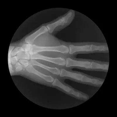
Fingers and wrist joints dislocation
Wrist Joint Dislocation
True dislocations of the radiocarpal joint (the main wrist joint) are relatively uncommon compared to fractures or finger dislocations [1]. Before the advent of X-rays, many wrist injuries mistakenly diagnosed as sprains were actually fractures, particularly of the distal radius (like a Colles' fracture) [1].
Isolated dislocation of the distal ulna (at the distal radioulnar joint - DRUJ) is also uncommon, occurring dorsally or volarly (towards the palm) [1].
Among dislocations involving the carpal (wrist) bones themselves, lunate dislocation (where the lunate bone displaces volarly) and perilunate dislocation (where the other carpal bones dislocate, usually dorsally, around a normally positioned lunate) are the most significant [1, 2]. These typically result from high-energy trauma, like a fall onto an outstretched hand [1, 2].
Closed reduction (non-surgical repositioning) of carpal dislocations, especially lunate dislocations, can be difficult and may fail due to interposed tissues or inherent instability [1, 2]. Surgical intervention (open reduction and internal fixation - ORIF) is often required to accurately restore alignment and stability [1, 2]. Failure to properly treat these complex injuries can lead to significant long-term wrist dysfunction, chronic pain, nerve compression (median nerve), and post-traumatic arthritis [1, 2].
Fingers Dislocation
Finger dislocations are common injuries, particularly in sports [1, 3]. Among these, dorsal dislocation of the thumb's metacarpophalangeal (MCP) joint (where the proximal phalanx displaces onto the back of the metacarpal head) is frequently observed [1, 3]. This injury typically results from a fall onto the hand or direct impact causing forced hyperextension of the thumb [1, 3].
During this injury, the volar plate (a thick ligament on the palm side of the joint) often ruptures, allowing the proximal phalanx to displace dorsally onto the metacarpal head [3].
Dislocations of the interphalangeal (IP) joints (proximal - PIP, and distal - DIP) of the fingers are even more common, often resulting from axial loading or hyperextension forces ("jammed finger") [1, 3]. Dorsal dislocations are the most frequent type for PIP and DIP joints as well [1, 3].
Video demonstrating the reduction technique for a thumb MCP joint dislocation.
Fingers Dislocation Symptoms and Reduction
For a dorsal thumb MCP dislocation, the thumb often appears shortened and is held in marked hyperextension at the MCP joint, with the interphalangeal (IP) joint possibly flexed, creating a 'bayonet' appearance [1, 3]. The base of the proximal phalanx may be palpable dorsally over the metacarpal, while the metacarpal head might be prominent on the volar (palm) side [1]. Attempts to move the joint are painful and restricted [1].
Closed reduction of a dorsal thumb MCP dislocation can sometimes be difficult if the metacarpal head becomes trapped between tendons (like the flexor pollicis longus), intrinsic muscles, or if the ruptured volar plate with its attached sesamoid bones becomes interposed in the joint (complex dislocation) [1, 3]. Simple dislocations, without entrapment, are usually easier to reduce [3].
Reduction often involves accentuating the hyperextension deformity to disengage any entrapped structures, followed by applying steady longitudinal traction and then flexing the proximal phalanx over the metacarpal head while applying volar pressure to its base [1, 3]. Adequate anesthesia (e.g., digital block or procedural sedation) is crucial [3]. If closed reduction attempts fail, open surgical reduction is necessary [1, 3].
Volar (palmar) dislocations of the thumb MCP joint are much rarer and typically result from forced hyperflexion [1]. Reduction involves traction, extension, and dorsal pressure on the base of the phalanx [1].
For PIP and DIP joint dislocations of the other fingers, the diagnosis is usually obvious from the visible deformity, finger alignment, and palpation of the displaced bone ends [1, 3]. X-rays are essential to confirm the dislocation type and rule out associated fractures [1, 3].
Reduction of PIP and DIP dislocations is typically achieved with longitudinal traction (pulling along the length of the finger) and gentle pressure applied to the base of the dislocated phalanx, guiding it back into place (e.g., gently flexing a dorsal dislocation after applying traction) [1, 3]. Local anesthesia (digital block) is usually sufficient [3]. After reduction, the joint's stability should be assessed, and the finger is typically splinted (often using buddy taping to an adjacent finger) for a few weeks, followed by range-of-motion exercises [1, 3].
Differential Diagnosis of Acute Finger/Wrist Injury
| Condition | Key Features / Distinguishing Points | Typical Investigations / Findings |
|---|---|---|
| Finger Dislocation (MCP/PIP/DIP) | Obvious deformity at the affected joint, inability to move the joint, pain, swelling. Often follows specific trauma (hyperextension, axial load). | X-ray confirms displacement of articular surfaces (e.g., phalanx base displaced relative to metacarpal/phalangeal head). Essential to rule out associated fracture. |
| Wrist (Carpal) Dislocation (Lunate/Perilunate) | Significant wrist pain, swelling, deformity, limited motion after high-energy trauma (fall on outstretched hand). May have median nerve symptoms (numbness/tingling). | X-ray (AP, lateral, oblique views) shows abnormal alignment of carpal bones (e.g., "spilled teacup" sign for lunate dislocation). CT often needed to fully define injury/fractures. |
| Distal Radius Fracture (e.g., Colles', Smith's) | Wrist pain, swelling, deformity ("dinner fork" in Colles'), tenderness over distal radius after fall. Carpal bones usually aligned with radius fragment. | X-ray shows fracture of the distal radius with characteristic displacement. |
| Scaphoid Fracture | Pain in anatomical snuffbox (base of thumb) after fall on outstretched hand. Swelling may be minimal. Often missed on initial X-rays. | Clinical suspicion high with snuffbox tenderness. Initial X-rays (including scaphoid view) may be negative. Repeat X-ray in 10-14 days, MRI, or CT may be needed to confirm. |
| Finger Fracture (Phalanx/Metacarpal) | Localized pain, swelling, tenderness, deformity, potential crepitus over the fracture site. May have rotational malalignment. Joint itself is aligned (unless fracture-dislocation). | X-ray confirms fracture line and displacement. |
| Ligament Sprain/Tear (e.g., Skier's Thumb - UCL) | Joint pain, swelling, instability, tenderness over the affected ligament. Occurs after specific mechanism (e.g., forced abduction of thumb for UCL tear). No gross deformity typical of dislocation. | Clinical exam shows laxity with stress testing (compare to contralateral side). X-rays normal or show small avulsion fracture. MRI confirms ligament tear. |
| Tendon Injury (e.g., Mallet Finger, Boutonniere, Extensor/Flexor Laceration) | Specific inability to extend or flex a joint actively. May have characteristic posture (e.g., DIP joint droop in Mallet Finger). Pain, swelling. Often associated with trauma or laceration. | Clinical exam demonstrates specific tendon function deficit. X-ray may show avulsion fracture (Mallet, Boutonniere) or be normal. Ultrasound/MRI can visualize tendon. |
References
- Skinner HB, McMahon PJ. Current Diagnosis & Treatment in Orthopedics. 5th ed. McGraw Hill; 2014. Chapter 8: Hand & Wrist Trauma.
- Rockwood CA, Green DP, Bucholz RW, Heckman JD. Rockwood and Green's Fractures in Adults. 8th ed. Lippincott Williams & Wilkins; 2014. Volume 1, Chapter 29: Carpal Dislocations and Instability.
- Nellans KW, Chung KC. Management of Common Finger Dislocations. Plast Reconstr Surg. 2013 Nov;132(5):810e-819e.
- Roberts DM, Khasriya R, Malone-Lee J. Hand Injuries: Dislocations. BMJ Clin Evid. 2011;2011:1110.
See also
- Achilles tendon inflammation (paratenonitis, ahillobursitis)
- Achilles tendon injury (sprain, rupture)
- Ankle and foot sprain
- Arthritis and arthrosis (osteoarthritis):
- Autoimmune connective tissue disease:
- Bunion (hallux valgus)
- Epicondylitis ("tennis elbow")
- Hygroma
- Joint ankylosis
- Joint contractures
- Joint dislocation:
- Knee joint (ligaments and meniscus) injury
- Metabolic bone disease:
- Myositis, fibromyalgia (muscle pain)
- Plantar fasciitis (heel spurs)
- Tenosynovitis (infectious, stenosing)
- Vitamin D and parathyroid hormone

