Hyperparathyroidism
Primary Hyperparathyroidism
Hyperparathyroidism, a condition of excessive parathyroid hormone (PTH) secretion, can occur at any age and affects both men and women, although it is more common in postmenopausal women [1]. Primary hyperparathyroidism refers to inappropriate, excessive PTH secretion originating from the parathyroid glands themselves, leading to hypercalcemia (high blood calcium) [1, 2].
The most common cause (80-85%) is a single parathyroid adenoma (a benign tumor) [1, 2]. Less often (10-15%), it results from hyperplasia involving multiple glands (usually all four) [1, 2]. Parathyroid carcinoma (cancer) is a rare cause (<1%) [1, 2]. Primary hyperparathyroidism can also be part of genetic syndromes like Multiple Endocrine Neoplasia (MEN) types 1 and 2A [1].
The diagnosis is primarily biochemical, established by demonstrating elevated or inappropriately normal serum PTH levels in the presence of hypercalcemia [1, 2]. Additional tests characterizing metabolic changes include measuring serum calcium (total and ionized), phosphate (often low or low-normal), albumin, vitamin D levels (25-hydroxyvitamin D), and 24-hour urine calcium (often high or normal) [1, 2]. Assessing renal phosphate clearance or tubular reabsorption of phosphate can provide further evidence of PTH action but is less commonly performed now [1].
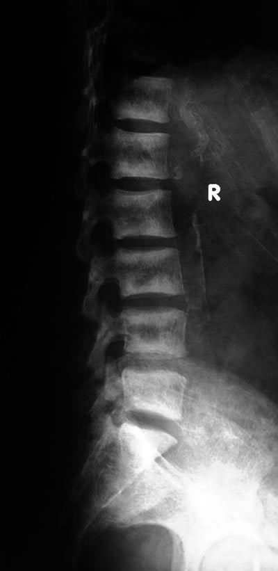
Untreated primary hyperparathyroidism increases the risk of complications related to chronic hypercalcemia and excessive PTH action, including osteoporosis (due to increased bone resorption), nephrolithiasis (kidney stones), nephrocalcinosis, bone pain, fractures, and potentially non-specific symptoms like fatigue, depression, constipation, and cognitive difficulties ("stones, bones, abdominal groans, thrones, and psychiatric overtones") [1, 2].
Secondary and Tertiary Hyperparathyroidism
Secondary hyperparathyroidism is a compensatory response by the parathyroid glands to chronic hypocalcemia (low blood calcium) or, less commonly, hyperphosphatemia [1, 2]. The glands enlarge (hyperplasia) and secrete more PTH in an attempt to normalize calcium levels [1]. The most common causes are:
- Chronic Kidney Disease (CKD): Impaired kidneys cannot adequately excrete phosphate (leading to hyperphosphatemia) and cannot efficiently activate vitamin D to its active form, 1,25-dihydroxyvitamin D (calcitriol) [1, 2]. Reduced calcitriol leads to decreased intestinal calcium absorption, causing hypocalcemia. Both hyperphosphatemia and hypocalcemia stimulate PTH secretion [1, 2].
- Vitamin D Deficiency: Insufficient dietary intake, lack of sun exposure, or malabsorption leads to inadequate calcium absorption from the gut, resulting in hypocalcemia and subsequent PTH elevation [1, 2].
In secondary hyperparathyroidism, serum calcium is typically low or normal, while PTH levels are elevated [1].
Tertiary hyperparathyroidism develops when the parathyroid glands, after prolonged stimulation in secondary hyperparathyroidism (usually due to end-stage renal disease), become autonomous and continue to oversecrete PTH even after the underlying cause of hypocalcemia is corrected (e.g., after successful kidney transplantation) [1, 2]. This results in hypercalcemia along with persistently elevated PTH levels [1, 2]. The glands are typically markedly hyperplastic, and sometimes an adenoma may develop within a hyperplastic gland [1].
Treatment for secondary hyperparathyroidism focuses on correcting the underlying cause (e.g., vitamin D replacement, phosphate binders, calcimimetics for CKD) [1, 2]. Tertiary hyperparathyroidism often requires parathyroidectomy (surgical removal of most of the parathyroid tissue) [1, 2].
Differential Diagnosis of Hypercalcemia
| Condition | Typical PTH Level | Key Features / Distinguishing Points |
|---|---|---|
| Primary Hyperparathyroidism | High or Inappropriately Normal | Most common cause of outpatient hypercalcemia. Often asymptomatic or mild symptoms ("stones, bones, groans, thrones, psychiatric overtones"). Low/normal phosphate, high urine calcium. Due to adenoma/hyperplasia. |
| Malignancy-Associated Hypercalcemia | Low (Suppressed) | Common cause of inpatient hypercalcemia. Often severe, rapid onset hypercalcemia with significant symptoms (confusion, dehydration). Due to PTHrP production by tumor (e.g., squamous cell, renal, breast) or bone metastases (e.g., multiple myeloma, breast). Underlying cancer often known or suspected. |
| Tertiary Hyperparathyroidism | High | Occurs in patients with long-standing secondary hyperparathyroidism (usually CKD) after correction of hypocalcemia (e.g., post-kidney transplant). Autonomous PTH secretion from hyperplastic glands. |
| Familial Hypocalciuric Hypercalcemia (FHH) | Normal or Mildly Elevated | Inherited disorder of calcium-sensing receptor. Lifelong mild-moderate asymptomatic hypercalcemia. Very low urine calcium excretion (low Ca/Cr clearance ratio). Usually benign. |
| Vitamin D Toxicity / Excess Calcitriol | Low (Suppressed) | Due to excessive vitamin D intake or granulomatous diseases producing calcitriol (e.g., sarcoidosis, TB, lymphoma). High serum 25(OH)D (toxicity) or 1,25(OH)2D (granulomatous dz). Phosphate often high. |
| Medications | Variable (Thiazides: often Mildly Elevated PTH; Lithium: often High/Normal PTH) | Thiazide diuretics reduce urine calcium excretion. Lithium affects parathyroid set point. Check medication history. |
| Hyperthyroidism | Low (Suppressed) | Mild hypercalcemia can occur due to increased bone turnover. Associated symptoms of thyrotoxicosis. |
| Immobilization | Low (Suppressed) | Prolonged bed rest increases bone resorption. Usually occurs in patients with high baseline bone turnover (e.g., adolescents, Paget's disease). |
| Milk-Alkali Syndrome | Low (Suppressed) | Excessive intake of calcium and absorbable alkali (e.g., calcium carbonate antacids). Associated with metabolic alkalosis and renal impairment. |
References
- Bilezikian JP, Khan A, Potts JT Jr, et al. Guidelines for the management of asymptomatic primary hyperparathyroidism: summary statement from the Fourth International Workshop. J Clin Endocrinol Metab. 2014 Oct;99(10):3561-9. (Or newer guidelines if available).
- Potts JT Jr. Diseases of the Parathyroid Glands and Other Hyper- and Hypocalcemic Disorders. In: Harrison's Principles of Internal Medicine. 20th ed. McGraw Hill; 2018. Chapter 424.
- Madani G, Lalam R, Trewhella M. The radiological manifestations of hyperparathyroidism. Clin Radiol. 2007 Sep;62(9):821-31.
See also
- Achilles tendon inflammation (paratenonitis, ahillobursitis)
- Achilles tendon injury (sprain, rupture)
- Ankle and foot sprain
- Arthritis and arthrosis (osteoarthritis):
- Autoimmune connective tissue disease:
- Bunion (hallux valgus)
- Epicondylitis ("tennis elbow")
- Hygroma
- Joint ankylosis
- Joint contractures
- Joint dislocation:
- Knee joint (ligaments and meniscus) injury
- Metabolic bone disease:
- Myositis, fibromyalgia (muscle pain)
- Plantar fasciitis (heel spurs)
- Tenosynovitis (infectious, stenosing)
- Vitamin D and parathyroid hormone


