Thoracic and lumbar injuries
Thoracic and lumbar injuries
The thoracic spine is often demonstrated well on the anteroposterior chest radiograph that forms part of the standard series of views requested in major trauma. This x ray may be the first to reveal an injury to the thoracic spine. Radiographs of the thoracic and lumbar spine must be specifically requested if a cervical spine injury has been sustained (because of the frequency with which injuries at more than one level coexist) or if signs of thoracic or lumbar trauma are detected when the patient is log rolled. In obtunded patients in whom the thoracic and lumbar spine cannot be evaluated clinically, the radiographs should be obtained routinely during the secondary survey or on admission to hospital. Unstable fractures of the pelvis are often associated with injuries to the lumbar spine.
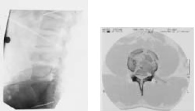
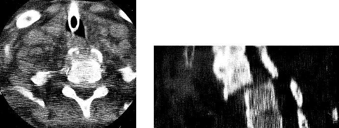
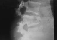
A significant force is normally required to damage the thoracic, lumbar, and sacral segments of the spinal cord, and the skeletal injury is usually evident on the standard anteroposterior and horizontal beam lateral radiographs. Burst fractures, and fractures affecting the posterior facet joints or pedicles, are unstable and more easily seen on the lateral radiograph. Instability requires at least two of the three columns of the spine to be disrupted. In simple wedge fractures, only the anterior column is disrupted and the injury remains stable. The demonstration of detail in the thoracic spine can be extremely difficult, particularly in the upper four vertebrae, and computed tomography (CT) is often required for better definition. Instability in thoracic spinal injuries may also be caused by sternal or bilateral rib fractures, as the anterior splinting effect of these structures will be lost.
A particular type of fracture, the Chance fracture, is typically found in the upper lumbar vertebrae. It runs transversely through the vertebral body and usually results from a shearing force exerted by the lap component of a seat belt during severe deceleration injury. These fractures are often associated with intra-abdominal or retroperitoneal injuries.
A haematoma in the posterior mediastinum is often seen around the thoracic fracture site, particularly in the anteroposterior view of the spine and sometimes on the chest radiograph requested in the primary survey. If there is any suspicion that these appearances might be due to traumatic aortic dissection, an arch aortogram will be required.
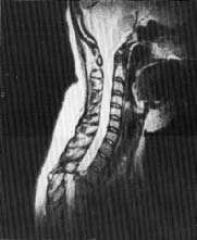
Fractures in the thoracic and lumbar spine are often complex and inadequately shown on plain films. CT demonstrates bony detail more accurately. MRI is used to demonstrate the extent of cord and soft tissue damage.
Indications for thoracic and lumbar radiographs:
- Major trauma
- Impaired consciousness
- Distracting injury
- Physical signs of thoracic or lumbar trauma
- Pelvic fractures
- Altered peripheral neurology
See also
- At the accident:
- History and epidemiology of spinal cord injury
- Spinal injuries management at the scene of the accident
- Evacuation and initial management at hospital:
- Evacuation and transfer to hospital of patients with spinal cord injuries
- Initial management of patients with spinal cord injuries at the receiving hospital
- Neurological assessment of patients with spinal cord injuries
- Spinal shock after severe spinal cord injury
- Partial spinal cord injury syndromes
- Radiological investigations:
- Initial radiography of patients with spinal cord injuries
- Cervical injuries
- Thoracic and lumbar injuries
- Early management and complications of spinal cord injuries — I:
- Respiratory complications
- Cardiovascular complications
- Prophylaxis against thromboembolism
- Initial bladder management
- The gastrointestinal tract
- Use of steroids and antibiotics
- The skin and pressure areas
- Care of the joints and limbs
- Later analgesia
- Trauma re-evaluation
- Early management and complications of spinal cord injuries — II:
- The anatomy of spinal cord injury
- The spinal injury (cervical, thoracic and lumbar spine)
- Transfer to a spinal injuries unit
- Medical management in the spinal injuries unit:
- The cervical spine injuries
- The cervicothoracic junction injuries
- Thoracic injuries
- Thoracolumbar and lumbar injuries
- Deep vein thrombosis and pulmonary embolism
- Autonomic dysreflexia
- Biochemical disturbances
- Para-articular heterotopic ossification
- Spasticity
- Contractures
- Pressure sores
- Urological management of patients with spinal cord injury:
- Nursing for people with spinal cord lesion:
- Physiotherapy after spinal cord injury:
- Respiratory management
- Mobilisation into a wheelchair
- Rehabilitation
- Recreation
- Incomplete lesions
- Children
- Occupational therapy after spinal cord injury:
- Hand and upper limb management
- Home resettlement
- Activities of daily living
- Communication
- Mobility
- Leisure
- Work
- Social needs of patient and family:
- Transfer of care from hospital to community:
- Education of patients
- Teaching the family and community staff
- Preparation for discharge from hospital
- Easing transfer from hospital to community
- Travel and holidays
- Follow-up
- Later management and complications after spinal cord injury — I:
- Late spinal instability and spinal deformity
- Pathological fractures
- Post-traumatic syringomyelia (syrinx, cystic myelopathy)
- Pain
- Sexual function
- Later management and complications after spinal cord injury — II:
- Later respiratory management of high tetraplegia
- Psychological factors
- The hand in tetraplegia
- Functional electrical stimulation
- Ageing with spinal cord injury
- Prognosis
- Spinal cord injury in the developing world:


