Medical management in the spinal injuries unit
Management of spinal cord injury in an acute specialised unit is associated with reduced mortality, increased neurological recovery, shorter length of stay and reduced cost of care, compared to treatment in a non-specialised centre. The objects of management are to prevent further spinal cord damage by appropriate reduction and stabilisation of the spine, to prevent secondary neuronal injury, and to prevent medical complications.
Objectives of medical management:
- Prevent further damage through reduction and immobilisation
- Prevent secondary neuronal injury
- Prevent medical complications
The cervical spine injuries
In injuries of the cervical spine skull traction is normally maintained for six weeks initially. The spine may be positioned in neutral or extension depending on the nature of the injury. Thus flexion injuries with suspected or obvious damage to the posterior ligamentous complex are treated by placing the neck in a degree of extension. The standard site of insertion of skull calipers need not be changed to achieve this; extension is achieved by correctly positioning a pillow or support under the shoulders. Most injuries are managed with the neck in the neutral position. An appropriately sized neck roll can also be inserted to maintain normal cervical lordosis and for the comfort of the patient.
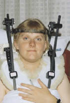
The application of a halo brace is a useful alternative to skull traction in many patients, once the neck is reduced. It provides stability and allows early mobilisation. Its use is often necessary for up to 12 weeks, when it can be replaced by a cervical collar if the neck is stable.
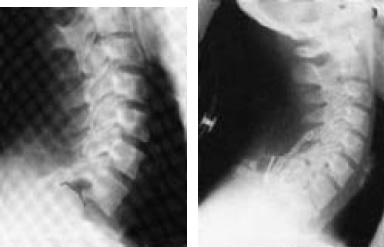
One of the most difficult aspects of cervical spine injury management is assessment of stability. Radiographs are taken regularly for position and at six weeks for evidence of bony union, immobilisation being continued for a further two to three weeks if there are any signs of instability. Once stability is achieved the patient is sat up in bed gradually during the course of a few days, wearing a firm cervical support such as a Philadelphia or Miami collar, before being mobilised into a wheelchair. This process is most conveniently achieved with a profiling bed, but the skin over the natal cleft and other pressure areas must be inspected frequently for signs of pressure or shearing. Some patients, particularly those with high level lesions, have postural hypotension when first mobilised because of their sympathetic paralysis, so profiling must not be hurried.
Cervical spine injuries:
- Skull traction for at least six weeks
- Halo traction (allows early mobilisation by conversion into halo brace in selected patients)
- Spinal fusion ( acute central disc prolapse (urgent decompression required), severe ligamentous damage, correction of major spinal deformity)
Antiembolism stockings and an abdominal binder help reduce the peripheral pooling of blood due to the sympathetic paralysis. Ephedrine 15–30 mg given 20 minutes before profiling starts is also effective. Once the spine is radiologically stable the firm collar can often be dispensed with at about 12 weeks after injury and a soft collar worn for comfort.
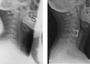
Twelve weeks after injury following plain x ray, if there is any likelihood of instability, flexion-extension radiography should be performed under medical supervision but if pain or paraesthesiae occur the procedure must be discontinued. It must be remembered that pain-induced muscle spasm may mask ligamentous injury and give a false sense of security. Most unstable injuries in the lower cervical spine are due to flexion or flexion-rotation forces and in the upper cervical spine to hyperextension. If internal fixation is indicated an anterior or posterior approach can be used, but if there is anterior cord compression, such as by a disc, anterior decompression and fixation is necessary. Fixation must be sound to avoid the need for extensive additional support.
Radiological signs of instability seen on standard lateral radiographs or flexion-extension views:
- Widening of gap between adjacent spinous processes
- Widening of intervertebral disc space
- Greater than 3.5 mm anterior or posterior displacement of vertebral body
- Increased angulation between adjacent vertebrae
The decision to perform spinal fusion is usually taken early, and sometimes it will have been performed in the district general hospital before transfer to the spinal injuries unit. The decision about when to operate will depend on the expertise and facilities available and the condition of the patient, but we suspect from our experience that early surgery in high lesion patients can sometimes precipitate respiratory failure, requiring prolonged ventilation. Some patients require late spinal fusion because of failed conservative treatment.
The upper cervical spine
As injuries of the upper cervical spine are often initially associated with acute respiratory failure, prompt appropriate treatment is important, including ventilation if necessary. Other patients may have little or no neurological deficit but again prompt treatment is important to prevent neurological deterioration.
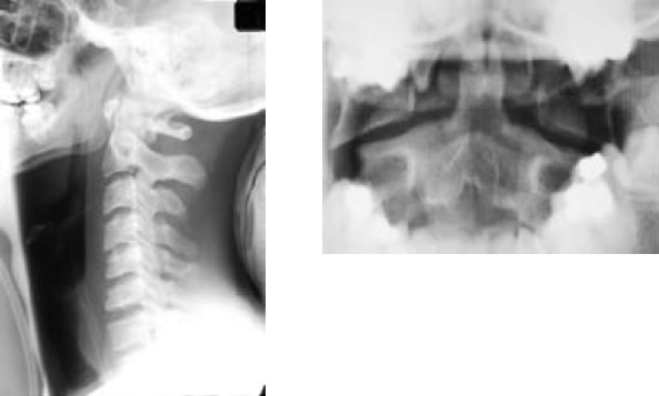
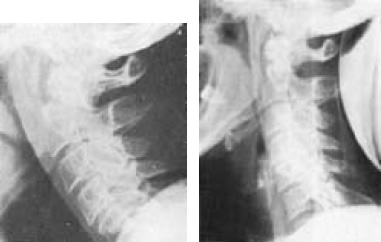
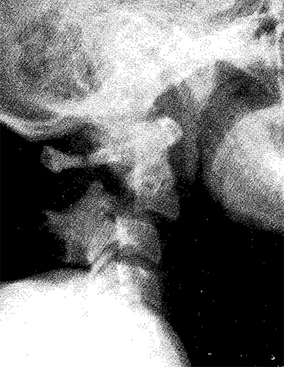
Fractures of the atlas are of two types. The most common, a fracture of the posterior arch, is due to an extension-compression force and is a stable injury which can be safely treated by immobilisation in a firm collar. The second type, the Jefferson fracture, is due to a vertical compression force to the vertex of the skull, resulting in the occipital condyles being driven downwards to produce a bursting injury, in which there is outward displacement of the lateral masses of the atlas and in which the transverse ligament may also have been ruptured. This is an unstable injury with the potential for atlanto-axial instability, and skull traction or immobilisation in a halo brace is necessary for at least eight weeks.
Fractures through the base of the odontoid process (type II fractures) are usually caused by hyperextension, and result in posterior displacement of the odontoid and posterior subluxation of Cl on C2; flexion injuries produce anterior displacement of the odontoid and anterior subluxation of Cl on C2. If displacement is considerable, reduction is achieved by gentle controlled skull traction under radiographic control. Immobilisation is continued for at least three to four months, depending on radiographic signs of healing. Halo bracing is very useful in managing this fracture. Atlanto-axial fusion may be undertaken by the anterior or posterior route if there is non-union and atlanto-axial instability. Anterior odontoid screw fixation may prevent rotational instability and avoid the need for a halo brace.
The "hangman’s" fracture, a traumatic spondylolisthesis of the axis, so called because the bony damage is similar to that seen in judicial hanging, is usually produced by hyperextension of the head on the neck, or less commonly with flexion. This results in a fracture through the pedicles of the axis in the region of the pars interarticularis, with an anterior slip of the C2 vertebral body on that of C3. Bony union occurs readily, but gentle skull traction should be maintained for six weeks, followed by immobilisation in a firm collar for a further two months. Great care must be taken to avoid overdistraction in this injury. Indeed, in all upper cervical fracture-dislocations once reduction has been achieved control can usually be obtained by reducing the traction force to only 1–2 kg. If more weight is used, neurological deterioration may result from overdistraction at the site of injury. An alternative approach when there is no bony displacement or when reduction has been achieved is to apply a halo brace. This avoids overdistraction from skull traction.
Ankylosing spondylitis predisposes the spine to fracture. It must be remembered that in this condition the neck is normally flexed, and to straighten the cervical spine will tend to cause respiratory obstruction, increase the deformity and risk further spinal cord damage. Help should be sought as soon as the problem is recognised.
The cervicothoracic junction injuries
Closed reduction of a C7–T1 facet dislocation is often difficult if not impossible, in which case operative reduction by facetectomy and posterior fusion is indicated, particularly in patients with an incomplete spinal cord lesion.
Thoracic injuries
The anatomy of the thoracic spine and the rib cage gives it added stability, although injuries to the upper thoracic spine are sometimes associated with a fracture of the sternum, which makes the injury unstable because of the loss of the normal anterior splinting effect of the sternum. It is very difficult to brace the upper thoracic spine, and if such a patient is mobilised too quickly a severe flexion deformity of the spine may develop.
In the majority of patients with a thoracic spinal cord injury, the neurological deficit is complete, and patients are usually managed conservatively by six to eight weeks’ bed rest.
Thoracolumbar and lumbar injuries
Most patients with thoracolumbar injuries can be managed conservatively with an initial period of bed rest for 8 to 12 weeks followed by gradual mobilisation in a spinal brace. If there is gross deformity or if the injury is unstable, especially if the spinal cord injury is incomplete, operative reduction, surgical instrumentation, and bone grafting correct the deformity and permit early mobilisation.
Isolated laminectomy has no place because it may render the spine unstable and does not achieve adequate decompression of the spinal cord except in the rare instance of a depressed fracture of a lamina. It must be combined with internal fixation and bone grafting. If spinal cord decompression is felt to be desirable, surgery should be aimed at the site of bony compression, which is generally anteriorly. An anterior approach with vertebrectomy by an experienced surgeon carries little added morbidity, except that it may cause significant deterioration in patients with pulmonary or chest wall injury. Dislocations and translocations can be dealt with by a posterior approach.
Before a patient with an unstable injury is mobilised, the spine is braced, the brace remaining in place until bony union occurs. Even if operative reduction has been undertaken, bracing may still be required for up to six months, depending on the type of spinal fusion performed.
Deep vein thrombosis and pulmonary embolism
Due to the very high incidence of thromboembolic complications, prophylaxis using antiembolism stockings and low molecular weight heparin should, in the absence of contraindications, be started within the first 72 hours of the accident. It is continued throughout the initial period of bed rest until the patient is fully mobile in a wheelchair and for a total of 8 weeks, or 12 weeks if there are additional risk factors such as a history of deep vein thrombosis, a lower limb fracture, or obesity.
An alternative is to commence warfarin as soon as the patient’s paralytic ileus has settled. If pulmonary embolism occurs the management is as for non-paralysed patients.
Autonomic dysreflexia
Autonomic dysreflexia is seen particularly in patients with cervical cord injuries above the sympathetic outflow but may also occur in those with high thoracic lesions above T6. It may occur at any time after the period of spinal shock and is usually due to a distended bladder caused by a blocked catheter, or to poor bladder emptying as a result of detrusor-sphincter dyssynergia. The distension of the bladder results in reflex sympathetic overactivity below the level of the spinal cord lesion, causing vasoconstriction and severe systemic hypertension. The carotid and aortic baroreceptors are stimulated and respond via the vasomotor centre with increased vagal tone and resulting bradycardia, but the peripheral vasodilatation that would normally have relieved the hypertension does not occur because stimuli cannot pass distally through the injured cord.
Characteristically the patient suffers a pounding headache, profuse sweating, and flushing or blotchiness of the skin above the level of the spinal cord lesion. Without prompt treatment, intracranial haemorrhage may occur.
Autonomic dysreflexia:
- Pounding headache
- Profuse sweating
- Flushing or blotchiness above level of lesion
- Danger of intracranial haemorrhage
Other conditions in which visceral stimulation can result in autonomic dysreflexia include urinary tract infection, bladder calculi, a loaded colon, an anal fissure, ejaculation during sexual intercourse, and labour.
Treatment consists of removing the precipitating cause. If this lies in the urinary tract catheterisation is often necessary. If hypertension persists nifedipine 5–10 mg sublingually, glyceryl trinitrate 300 micrograms sublingually, or phentolamine 5–10 mg intravenously is given. If inadequately treated the patient can become sensitised and develop repeated attacks with minimal stimuli. Occasionally the sympathetic reflex activity may have to be blocked by a spinal or epidural anaesthetic. Later management may include removal of bladder calculi or sphincterotomy if detrusor-sphincter dyssynergia is causing the symptoms; performed under spinal anaesthesia, the risk of autonomic dysreflexia is lessened.
Treatment of autonomic dysreflexia:
- Remove the cause
- Sit patient up
- Treat with Nifedipine 5–10 mg capsule (bite and swallow) or Glyceryl trinitrate 300 µg sublingually. If blood pressure continues to rise despite intervention, treat with antihypertensive drug e.g. phentolamine 5–10 mg intravenously in 2.5 mg increments
- Spinal or epidural anaesthetic (rarely)
Biochemical disturbances
Hyponatraemia
The aetiology of hyponatraemia is multifactorial, involving fluid overload, diuretic usage, the sodium depleting effects of drugs such as carbamazepine, and inappropriate antidiuretic hormone secretion.
It may occur (1) during the acute stage of spinal cord injury, when the patient is on intravenous fluids, or (2) in the chronic phase, often in association with systemic sepsis frequently of chest or urinary tract origin, and often exacerbated by the patient increasing their oral fluid intake in an attempt to eradicate a suspected urinary infection.
Treatment depends on the severity and the cause. Sepsis should be controlled, fluids restricted, and medication reviewed. Hypertonic saline (2N) should be avoided because of the risk of central pontine myelinolysis. Furosemide (frusemide) and potassium supplements are useful, but the rate of correction of the serum sodium must be managed carefully.
Occasionally hyponatraemia is prolonged and in this situation demeclocycline hydrochloride is useful.
Hypercalcaemia
Any prolonged period of immobility results in the mobilisation of calcium from the bones, and, particularly in tetraplegics, this can be associated with symptomatic hypercalcaemia. The diagnosis is often difficult, and symptoms can include constipation, abdominal pain, and headaches. The problem is uncommon and diagnosis may be delayed, if the serum calcium is not measured.
Treatment involves hydration, achieving a diuresis (with a fluid load and furosemide (frusemide)), and the use of oral sodium etidronate or intravenous disodium pamidronate. Once the patient is fully mobile the problem usually resolves.
Biochemical disturbances:
| Hyponatraemia | Hypercalcaemia | ||
| Acute Hyponatraemia | - due to excessive intravenous fluids | Hypercalcaemia Symptoms | - constipation |
| Chronic Hyponatraemia | - systemic sepsis - excessive oral fluid intake - drug induced e.g. carbamazepine |
Hypercalcaemia Treatment | - hydration - achieve diuresis - oral disodium etidronate or intravenous disodium pamidronate |
| Hyponatraemia Treatment | - treat sepsis - control fluid intake - review drugs - furosemide, potassium supplements - demeclocycline (occasionally) |
||
Para-articular heterotopic ossification
After injury to the spinal cord new bone is often laid down in the soft tissues around paralysed joints, particularly the hip and knee. The cause is unknown, although local trauma has been suggested. It usually presents with erythema, induration, or swelling near a joint. There is pronounced osteoblastic activity, but the new bone formed does not mature for at least 18 months. This has an important bearing on treatment in that if excision of heterotopic bone is required because of gross restriction of movement or bony ankylosis of a joint, surgery is best delayed for at least 18 months — until the new bone is mature. Earlier surgical intervention may provoke further new bone formation, thus compounding the original condition. Treatment with disodium etidronate suppresses the mineralisation of osteoid tissue and may reverse up to half of early lesions when used for 3–6 months, and non-steroidal anti-inflammatory drugs are also used to prevent the progression of this complication. Postoperative radiotherapy may halt the recurrence of the problem if early surgical intervention has to be performed.
Spasticity
Spasticity is seen only in patients with upper motor neurone lesions of the cord whose intact spinal reflex arcs below the level of the lesion are isolated from higher centres. It usually increases in severity during the first few weeks after injury, after the period of spinal shock. In incomplete lesions it is often more pronounced and can be severe enough to prevent patients with good power in the legs from walking. Patients with severe spasticity and imbalance of opposing muscle groups have a tendency to develop contractures. It is important to realise that once a contracture occurs spasticity is increased and a vicious circle is established with further deformity resulting. Although excessive spasticity may hamper patients’ activities or even throw them out of their wheelchairs or make walking impossible, spasticity may have advantages. It maintains muscle bulk and possibly bone density, and improves venous return.
Factors that aggravate spasticity:
- Urinary tract infection or calculus
- Infected ingrowing toenail
- Pressure sores
- Anal fissure
- Fracture
- Contractures
Treatment of severe spasticity is indicated if it interferes with activities of daily living, and is initially directed at removing any obvious precipitating cause. An irritative lesion in the paralysed part, such as a pressure sore, urinary tract infection or calculus, anal fissure, infected ingrowing toenail, or fracture, tends to increase spasticity.
Passive stretching of spastic muscles and regular standing are helpful in relieving spasticity and preventing contractures. The drugs most commonly used to decrease spasticity are baclofen and tizanidine, which act at spinal level, and dantrolene sodium, which acts directly on skeletal muscle. Although diazepam relieves spasticity, its sedative action and habit-forming tendency limit its usefulness. With these drugs, the liver function tests need to be closely monitored.
If spasticity is localised it can be relieved by interrupting the nerve supply to the muscles affected by neurectomy after a diagnostic block with a long-acting local anaesthetic (bupivacaine). For example, in patients with severe hip adductor spasticity obturator neurectomy is effective. Alternatively, motor point injections, initially with bupivacaine, followed by either 6% aqueous phenol or 45% ethyl alcohol for a more lasting effect, are useful in selected patients. Botulinum toxin also has a limited use in patients with localised spasticity.
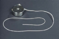
If oral agents are failing to control generalised spasticity intrathecal baclofen will often provide relief. If a small test dose of 50 micrograms baclofen given by lumbar puncture relieves the spasticity, a reservoir and pump can be implanted to provide regular and long-term delivery of the drug. It is now rare to have to resort to destructive procedures involving surgical or chemical neurectomy, or intrathecal blocks with 6% aqueous phenol or absolute alcohol. The effect of phenol usually lasts a few months, that of alcohol is permanent. The main disadvantage in the use of either is that they convert an upper motor neurone to a lower motor neurone lesion and thus affect bladder, bowel, and sexual function.
Management of spasticity:
- Treat factors that aggravate spasticity
- Improve comfort/posture
- Manage pain
- Passively stretch spastic muscles
- Regular standing
- Oral therapy (baclofen, tizanidine, dantrolene, diazepam)
- Botulinum toxin
- Motor point injections
- Implanted drug delivery system for administration of intrathecal baclofen
If the above fail, are contraindicated, or are unavailable:
- tendon release and/or neurectomy, and other orthopaedic procedures
- intrathecal block (rarely used) (6% aqueous phenol, absolute alcohol)
Contractures
A contracture may be a result of immobilisation, spasticity, or muscle imbalance between opposing muscle groups. It may respond to conservative measures such as gradual stretching of affected muscles, often with the use of splints. If these measures fail to correct the deformity or are inappropriate, then surgical correction by tenotomy, tendon lengthening, or muscle division may be required. For example, a flexion contracture of the hip responds to an iliopsoas myotomy with division of the anterior capsule and soft tissues over the front of the joint.
Treatment of contractures:
- Gradual stretching ± splints
- Tenotomy
- Tendon lengthening
- Muscle and soft tissue division
Prevention of pressure sores:
- Regular relief of pressure
- Regular checking of skin, using mirror
- Avoid all pressure if red mark develops
- Suitable cushion and mattress, checked regularly
- Avoid tight clothes and hard seams
Pressure sores
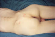
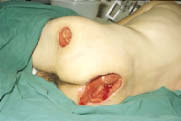
Pressure sores form as a result of ischaemia, caused by unrelieved pressure, particularly over bony prominences. They may affect not only the skin but also subcutaneous fat, muscle, and deeper structures. If near a joint, septic arthritis may supervene. The commonest sites are over the ischial tuberosity, greater trochanter, and sacrum. Pressure sores are a major cause of readmission to hospital, yet they are generally preventable by vigilance and recognition of simple principles.
Regular changes of position in bed every two to three hours and lifting in the wheelchair every 15 minutes are essential. A suitable mattress and wheelchair cushion are particularly important. The cushion should be selected for the individual patient after measuring the interface pressures between the ischial tuberosities and the cushion. Cushions need frequent checking and renewing if necessary. Shearing forces to the skin from underlying structures are avoided by correct lifting; the skin should never be dragged along supporting surfaces. Patients must not lie for long periods with the skin unprotected on x ray diagnostic units or on operating tables (in this situation Roho mattress sections placed under the patient are of benefit). A pressure clinic is extremely useful in checking the sitting posture, assessing the wheelchair and cushion, and generally instilling pressure consciousness into patients. If a red mark on the skin is noticed which does not fade within 20 minutes the patient should avoid all pressure on that area until the redness and any underlying induration disappears.
If an established sore is present, any slough is excised and the wound is dressed with a desloughing agent if necessary. Once the wound is clean and has healthy granulation tissue, occlusive dressings may be used. Complete relief of pressure on the affected area is essential until healing has occurred. Indications for surgery are: (1) a large sore which would take too long to heal using conservative methods; (2) a sore with infected bone in its base; (3) a discharging sinus with an underlying bursa. If possible, surgical treatment is by excision of the sore and any underlying bony prominence, with direct closure in layers, leaving a small linear scar. Most pressure sores can be managed in this way. Recurrence is uncommon and if it occurs can be more easily treated after this type of surgery than if large areas of tissues have been disturbed by previous use of a flap.
Treatment of pressure sores:
- Conservative — complete relief of pressure, if slough, treat with desloughing agent or excise, treat general condition, e.g. correct anaemia
- Surgical — direct closure if possible, with removal of underlying bony prominence
See also
- At the accident:
- History and epidemiology of spinal cord injury
- Spinal injuries management at the scene of the accident
- Evacuation and initial management at hospital:
- Evacuation and transfer to hospital of patients with spinal cord injuries
- Initial management of patients with spinal cord injuries at the receiving hospital
- Neurological assessment of patients with spinal cord injuries
- Spinal shock after severe spinal cord injury
- Partial spinal cord injury syndromes
- Radiological investigations:
- Initial radiography of patients with spinal cord injuries
- Cervical injuries
- Thoracic and lumbar injuries
- Early management and complications of spinal cord injuries — I:
- Respiratory complications
- Cardiovascular complications
- Prophylaxis against thromboembolism
- Initial bladder management
- The gastrointestinal tract
- Use of steroids and antibiotics
- The skin and pressure areas
- Care of the joints and limbs
- Later analgesia
- Trauma re-evaluation
- Early management and complications of spinal cord injuries — II:
- The anatomy of spinal cord injury
- The spinal injury (cervical, thoracic and lumbar spine)
- Transfer to a spinal injuries unit
- Medical management in the spinal injuries unit:
- The cervical spine injuries
- The cervicothoracic junction injuries
- Thoracic injuries
- Thoracolumbar and lumbar injuries
- Deep vein thrombosis and pulmonary embolism
- Autonomic dysreflexia
- Biochemical disturbances
- Para-articular heterotopic ossification
- Spasticity
- Contractures
- Pressure sores
- Urological management of patients with spinal cord injury:
- Nursing for people with spinal cord lesion:
- Physiotherapy after spinal cord injury:
- Respiratory management
- Mobilisation into a wheelchair
- Rehabilitation
- Recreation
- Incomplete lesions
- Children
- Occupational therapy after spinal cord injury:
- Hand and upper limb management
- Home resettlement
- Activities of daily living
- Communication
- Mobility
- Leisure
- Work
- Social needs of patient and family:
- Transfer of care from hospital to community:
- Education of patients
- Teaching the family and community staff
- Preparation for discharge from hospital
- Easing transfer from hospital to community
- Travel and holidays
- Follow-up
- Later management and complications after spinal cord injury — I:
- Late spinal instability and spinal deformity
- Pathological fractures
- Post-traumatic syringomyelia (syrinx, cystic myelopathy)
- Pain
- Sexual function
- Later management and complications after spinal cord injury — II:
- Later respiratory management of high tetraplegia
- Psychological factors
- The hand in tetraplegia
- Functional electrical stimulation
- Ageing with spinal cord injury
- Prognosis
- Spinal cord injury in the developing world:


