Initial radiography of patients with spinal cord injuries
Initial radiography of patients with spinal cord injuries
Radiological investigation of a high standard is crucial to the diagnosis of a spinal injury. Initial radiographs are taken in the emergency department. Most emergency departments rely on the use of mobile radiographic equipment for investigating seriously ill patients, but the quality of films obtained in this way is usually inferior.
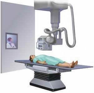
Once the patient’s condition is stable, radiographs can be taken in the radiology department. In the presence of neurological symptoms, a doctor should be in attendance to ensure that any spinal movement is minimised. Sandbags and collars are not always radiolucent, and clearer radiographs may be obtained if these are removed after preliminary films have been taken. Plain x ray pictures in the lateral and anteroposterior projections are fundamental in the diagnosis of spinal injuries. Special views, computed tomography (CT), and magnetic resonance imaging (MRI) are used for further evaluation.
Spinal cord injury without radiological abnormality (SCIWORA) may occur due to central disc prolapse, ligamentous damage, or cervical spondylosis which narrows the spinal canal, makes it more rigid, and therefore renders the spinal cord more vulnerable to trauma (particularly in cervical hyperextension injuries). SCIWORA is also relatively common in injured children because greater mobility of the developing spine affords less protection to the spinal cord.
See also
- At the accident:
- History and epidemiology of spinal cord injury
- Spinal injuries management at the scene of the accident
- Evacuation and initial management at hospital:
- Evacuation and transfer to hospital of patients with spinal cord injuries
- Initial management of patients with spinal cord injuries at the receiving hospital
- Neurological assessment of patients with spinal cord injuries
- Spinal shock after severe spinal cord injury
- Partial spinal cord injury syndromes
- Radiological investigations:
- Initial radiography of patients with spinal cord injuries
- Cervical injuries
- Thoracic and lumbar injuries
- Early management and complications of spinal cord injuries — I:
- Respiratory complications
- Cardiovascular complications
- Prophylaxis against thromboembolism
- Initial bladder management
- The gastrointestinal tract
- Use of steroids and antibiotics
- The skin and pressure areas
- Care of the joints and limbs
- Later analgesia
- Trauma re-evaluation
- Early management and complications of spinal cord injuries — II:
- The anatomy of spinal cord injury
- The spinal injury (cervical, thoracic and lumbar spine)
- Transfer to a spinal injuries unit
- Medical management in the spinal injuries unit:
- The cervical spine injuries
- The cervicothoracic junction injuries
- Thoracic injuries
- Thoracolumbar and lumbar injuries
- Deep vein thrombosis and pulmonary embolism
- Autonomic dysreflexia
- Biochemical disturbances
- Para-articular heterotopic ossification
- Spasticity
- Contractures
- Pressure sores
- Urological management of patients with spinal cord injury:
- Nursing for people with spinal cord lesion:
- Physiotherapy after spinal cord injury:
- Respiratory management
- Mobilisation into a wheelchair
- Rehabilitation
- Recreation
- Incomplete lesions
- Children
- Occupational therapy after spinal cord injury:
- Hand and upper limb management
- Home resettlement
- Activities of daily living
- Communication
- Mobility
- Leisure
- Work
- Social needs of patient and family:
- Transfer of care from hospital to community:
- Education of patients
- Teaching the family and community staff
- Preparation for discharge from hospital
- Easing transfer from hospital to community
- Travel and holidays
- Follow-up
- Later management and complications after spinal cord injury — I:
- Late spinal instability and spinal deformity
- Pathological fractures
- Post-traumatic syringomyelia (syrinx, cystic myelopathy)
- Pain
- Sexual function
- Later management and complications after spinal cord injury — II:
- Later respiratory management of high tetraplegia
- Psychological factors
- The hand in tetraplegia
- Functional electrical stimulation
- Ageing with spinal cord injury
- Prognosis
- Spinal cord injury in the developing world:

