Changes of the nasal mucosa in influenza, diphtheria, measles and scarlet fever
- Nasal Mucosal Changes in Influenza (Flu)
- Nasal Manifestations of Diphtheria
- Nasal Involvement in Measles (Rubeola)
- Nasal Mucosal Changes in Scarlet Fever
- Differential Diagnosis of Rhinitis in Febrile Exanthematous Illnesses
- General Management Principles for Nasal Symptoms in Systemic Infections
- When to Consult an ENT Specialist or Physician
- References
Nasal Mucosal Changes in Influenza (Flu)
Influenza, commonly known as the flu, is an acute viral infection of the respiratory tract caused by influenza viruses (Types A, B, and C). Viral infections like influenza frequently cause significant inflammatory changes not only in the nasal passages (rhinitis) but also in the paranasal sinuses (sinusitis). The presentation and severity can vary, especially with age.
Pathophysiology and Clinical Features
Influenza virus invades the respiratory epithelium, leading to cell damage, inflammation, and increased vascular permeability. This results in characteristic nasal symptoms:
- Nasal Discharge: Initially, the rhinitis associated with influenza is characterized by abundant serous (clear, watery) discharge. This can progress to mucoid, mucopurulent, or even purulent discharge if secondary bacterial infection occurs. Occasionally, the discharge may be blood-tinged due to mucosal friability.
- Nasal Congestion: Significant swelling of the nasal mucosa leads to nasal obstruction.
- Mucosal Hemorrhages: Small hemorrhages (petechiae or ecchymoses) are often visible on the nasal mucous membrane due to viral damage to capillaries and intense inflammation.
- Systemic Symptoms: Influenza typically presents with more severe systemic toxic effects than common acute respiratory diseases (colds). These include abrupt onset of high fever, chills, headache, myalgia (muscle aches), malaise, and fatigue.
- In **infants**, the flu may begin atypically, sometimes with a loss of body weight as an early sign. The temperature response can be variable, sometimes remaining subfebrile.
- In **children aged 1 to 4 years**, the febrile response is generally more pronounced than in infants under 1 year old.
- Complications: Acute otitis media (middle ear infection) is a common complication, especially in infants and young children, due to Eustachian tube dysfunction secondary to nasopharyngeal inflammation. Sinusitis is also a frequent sequela. More severe complications include viral or secondary bacterial pneumonia.
Management and Prevention
The treatment of influenza and its associated nasal symptoms is primarily supportive and is often managed in conjunction with an infectious disease specialist or primary care physician, especially in severe cases or high-risk individuals.
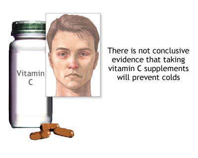
- Supportive Care: Bed rest, adequate hydration, and rational nutrition rich in vitamins (though the role of high-dose vitamin C in preventing or treating flu is a misconception; balanced nutrition is key).
- Symptomatic Relief:
- Antipyretics/analgesics (e.g., acetaminophen, ibuprofen) for fever and pain.
- Nasal saline sprays or drops to help clear secretions and soothe mucosa.
- In infants, gentle nasal aspiration before feeding can facilitate breathing during sucking.
- Antiviral Medications: Prescription antiviral drugs (e.g., oseltamivir, zanamivir, baloxavir) can shorten the duration and severity of influenza if started early (typically within 48 hours of symptom onset), especially for high-risk individuals or severe illness.
- Management of Complications: Antibiotics are indicated only if secondary bacterial infection (e.g., bacterial sinusitis, otitis media, pneumonia) is diagnosed.
- Prevention: Annual influenza vaccination is the most effective way to prevent influenza and its complications. Good hand hygiene and respiratory etiquette are also important.
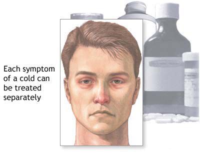
The rest of the local nasal treatment is generally similar to that for a common (banal) acute rhinitis, focusing on symptom relief and maintaining nasal hygiene.
Nasal Manifestations of Diphtheria
Diphtheria is a serious bacterial infection caused by toxin-producing strains of *Corynebacterium diphtheriae*. While its incidence has dramatically decreased in regions with widespread vaccination, it remains a concern in undervaccinated populations. Nasal diphtheria is one of the clinical forms of the disease.
Characteristics of Nasal Diphtheria
Nasal diphtheria typically presents with:
- Formation of Pseudomembranes: The hallmark of diphtheria is the formation of adherent, grayish-white or yellowish pseudomembranes on the mucosal surfaces. In nasal diphtheria, these films are found in the nasal cavity, often on the septum and turbinates. Attempts to remove the membrane usually cause bleeding.
- Nasal Discharge: Often initially serosanguineous (watery and blood-tinged), later becoming mucopurulent and sometimes foul-smelling. Unilateral discharge is common.
- Excoriation of Nares: The irritant discharge can cause excoriation and crusting around the nostrils and upper lip.
- Regional Lymphadenopathy: Swelling of cervical lymph nodes may occur, though typically less pronounced than in pharyngeal diphtheria ("bull neck").
- Systemic Intoxication: While often milder than pharyngeal or laryngeal diphtheria, systemic effects from diphtheria toxin (e.g., malaise, low-grade fever, impaired general condition of the child) can still be present, particularly if toxin absorption is significant or if there is concurrent involvement of other sites.
It is important to note that atypical forms of diphtheria can occur, sometimes presenting with poorly expressed signs, making diagnosis challenging. Differential diagnosis includes fibrinous rhinitis of non-diphtheritic origin (e.g., due to other bacteria like Streptococcus) and adenoviral diseases with predominant nasal localization.
Diagnosis and Treatment Imperatives
The diagnosis of diphtheria is based on:
- Clinical Suspicion: Presence of pseudomembranes, characteristic discharge, and compatible systemic symptoms.
- Bacteriological Studies: Culture of swabs taken from beneath the edge of the pseudomembrane on special media (e.g., Loeffler's or Tinsdale's agar) to isolate *C. diphtheriae*. Toxin production by the isolate must be confirmed (e.g., Elek test or PCR for tox gene).
- Epidemiological Data: History of exposure or lack of immunization.
Treatment of diphtheria is a medical emergency and must be carried out in an infectious disease hospital setting:
- Diphtheria Antitoxin (DAT): The cornerstone of treatment is the prompt parenteral administration of diphtheria antitoxin to neutralize circulating toxin. Dosage depends on the severity and duration of illness.
- Antibiotics: Antibiotics (e.g., penicillin or erythromycin) are given to eradicate *C. diphtheriae*, stop further toxin production, and prevent spread. They are an adjunct to antitoxin, not a substitute.
- Supportive Care: Including airway management if obstruction is present (rare in isolated nasal diphtheria but possible with pharyngeal spread), bed rest, and nutritional support. For infants, expressed breast milk is important.
- Local Nasal Care: To aid in the rejection and removal of films and excess mucus, gentle alkaline inhalations (e.g., 1-2% sodium bicarbonate solution) may be used. Historically, local application of a 2% boric acid solution with adrenaline was used to cleanse the nasal cavity, but modern practice emphasizes gentle suctioning and saline.
Prevention of diphtheria relies on widespread childhood immunization with diphtheria toxoid-containing vaccines (e.g., DTaP, Tdap, Td). Isolation of sick individuals, identification and treatment of carriers, and prophylactic measures (vaccination and sometimes antibiotics) for close contacts are crucial public health measures.
Nasal Involvement in Measles (Rubeola)
Measles is a highly contagious viral illness caused by the measles virus (a paramyxovirus). It is characterized by a prodromal phase of fever, malaise, cough, coryza (rhinitis), and conjunctivitis, followed by the appearance of a maculopapular rash.
Prodromal Symptoms, Rhinitis, and Koplik's Spots
Inflammatory processes in the nasal cavity (rhinitis) are a prominent feature of the prodromal stage of measles, typically appearing 2-4 days before the onset of the characteristic skin rash. This "measles rhinitis" usually accompanies the other "3 Cs" of the prodrome: cough and conjunctivitis. The rhinitis persists until the skin rash begins to fade.
Nasal symptoms include:
- Profuse Nasal Discharge: Initially serous, then becoming more mucoid or mucopurulent. The discharge may sometimes be tinged with blood.
- Nasal Congestion and Sneezing.
- Inflamed Nasal Mucosa: Appears red and swollen on examination.
A pathognomonic sign of measles, appearing 1-2 days before the skin rash, is **Koplik's spots**. These are small, bluish-white spots resembling grains of salt on an erythematous base, typically found on the buccal mucosa opposite the molar teeth. While not directly in the nose, their presence strongly supports a measles diagnosis when nasal symptoms are also present.
Inflammation in measles can extend beyond the nasal cavity to involve the paranasal sinuses (sinusitis), larynx (laryngitis, croup), trachea (tracheitis), and bronchi (bronchitis). Conjunctivitis ("pink eye") is also very common. Acute otitis media is a frequent complication, particularly in young children.
Management and Complications
The diagnosis of measles is usually based on the typical clinical presentation (prodrome, Koplik's spots, characteristic rash progression), especially during an outbreak. Laboratory confirmation can be done by detecting measles-specific IgM antibodies in serum or measles RNA by RT-PCR from respiratory secretions or urine.
Treatment for measles is primarily supportive, similar to that for common viral rhinitis or other viral URIs, as there is no specific antiviral therapy for uncomplicated measles:
- Rest and hydration.
- Antipyretics for fever.
- Humidified air for cough and nasal congestion.
- Nutritional support.
- Vitamin A supplementation is recommended by the WHO for all children with measles, especially in areas with vitamin A deficiency, as it can reduce morbidity and mortality.
Complications of measles can be serious and include otitis media, pneumonia (viral or secondary bacterial), encephalitis, laryngotracheobronchitis (croup), diarrhea, and, rarely, subacute sclerosing panencephalitis (SSPE) years later. Bacterial superinfections require antibiotic treatment.
Prevention through widespread measles vaccination (MMR or MMRV vaccine) is highly effective and the cornerstone of public health efforts to control the disease.
Nasal Mucosal Changes in Scarlet Fever
Scarlet fever is a bacterial illness caused by toxin-producing strains of *Streptococcus pyogenes* (Group A Streptococcus), the same bacteria that cause strep throat. It is characterized by a sore throat, fever, and a distinctive sandpaper-like rash.
Rhinitis as Part of Streptococcal Infection
While the primary site of infection in scarlet fever is usually the pharynx (strep throat), changes in the nasal mucous membrane can occur, although they are not typically specific or defining features of the disease itself. Nasal involvement in scarlet fever is generally similar to that seen with common banal rhinitis or the rhinitis accompanying other acute febrile illnesses.
Symptoms may include:
- Mild to moderate nasal congestion.
- Nasal discharge, which can be serous, mucoid, or mucopurulent.
- Inflamed and hyperemic (red) nasal mucosa.
These nasal symptoms are part of the broader upper respiratory tract inflammation caused by the streptococcal infection and its toxins. They are usually overshadowed by the more prominent symptoms of pharyngitis, fever, and the characteristic rash.
Diagnosis and Treatment
The diagnosis of scarlet fever typically does not present major difficulties, especially when the characteristic rash is present, along with other signs such as a "strawberry tongue," exudative tonsillitis (specific tonsillitis), and evidence of toxemia (systemic effects of bacterial toxins). A rapid strep test or throat culture confirms Group A Streptococcus infection.
Treatment for scarlet fever is carried out in an appropriate setting, which may include an infectious disease department for more severe cases, and primarily involves:
- Antibiotics: Penicillin (or amoxicillin) is the drug of choice to eradicate the streptococcal infection, prevent complications like rheumatic fever, and reduce contagiousness. For penicillin-allergic individuals, alternatives like macrolides or cephalosporins are used. A full course of antibiotics is essential.
- Supportive Care: Rest, hydration, and antipyretics/analgesics for fever and pain.
- Local Throat Care: Gargling with saline or mild antiseptic solutions (e.g., historically, 3% boric acid solution) may provide symptomatic relief for the sore throat.
- Antitoxin (Historical): Polyvalent anti-streptococcal serum was used in the past for severe toxic cases but is not part of standard modern therapy.
Nasal symptoms are managed symptomatically as part of the overall treatment for the streptococcal infection.
Differential Diagnosis of Rhinitis in Febrile Exanthematous Illnesses
Several infectious diseases present with fever, rash (exanthem), and rhinitis. Differentiating them based on nasal and other clinical features is important:
| Disease | Nasal Symptoms | Key Differentiating Features |
|---|---|---|
| Influenza | Serous to mucopurulent discharge, congestion, possible mucosal hemorrhages. | Abrupt onset, high fever, myalgia, headache, cough. Rash is not typical unless a co-infection or drug reaction occurs. |
| Nasal Diphtheria | Serosanguineous or mucopurulent discharge (often unilateral), excoriation of nares, grayish pseudomembranes in nose. | Low-grade fever, malaise. Membrane bleeds on removal. Culture positive for *C. diphtheriae*. |
| Measles (Rubeola) | Profuse coryza (serous/mucoid discharge), congestion, sneezing in prodrome. | Prodrome of fever, cough, conjunctivitis. Koplik's spots on buccal mucosa. Maculopapular rash starting on face and spreading downwards. |
| Scarlet Fever | Non-specific rhinitis (congestion, mucoid/mucopurulent discharge). | Sore throat (strep pharyngitis), fever, "strawberry tongue," fine sandpaper-like rash prominent in skin folds (Pastia's lines). Positive strep test/culture. |
| Rubella (German Measles) | Mild coryza and conjunctivitis may occur in prodrome. | Milder illness than measles. Maculopapular rash (less confluent), postauricular/suboccipital lymphadenopathy. Forchheimer spots on soft palate. |
| Varicella (Chickenpox) | Vesicular lesions may rarely occur on nasal mucosa. General URI symptoms possible. | Pruritic vesicular rash in crops, fever, malaise. |
| Common Cold (Viral URI) | Serous to mucopurulent discharge, congestion, sneezing. | Milder systemic symptoms, lower fever. No characteristic rash or specific oral lesions like Koplik's spots. |
General Management Principles for Nasal Symptoms in Systemic Infections
While specific treatment targets the underlying infectious agent (e.g., antivirals for influenza, antitoxin and antibiotics for diphtheria, antibiotics for scarlet fever), management of the associated nasal symptoms is generally supportive and aims to:
- Maintain Nasal Patency: Gentle suctioning (especially in infants), saline nasal drops or sprays to loosen secretions. Short-term use of topical decongestants may be considered for severe congestion interfering with feeding or sleep, but with caution regarding duration.
- Soothe Mucosal Irritation: Humidified air can help. Adequate hydration thins secretions.
- Control Fever and Pain: Antipyretics/analgesics like acetaminophen or ibuprofen.
- Prevent Complications: Monitor for signs of secondary bacterial infections like otitis media or sinusitis, which may require specific antibiotic treatment.
Addressing the primary systemic infection is paramount for resolving the associated nasal manifestations.
When to Consult an ENT Specialist or Physician
Consultation with a physician or an ENT specialist is important if:
- Nasal symptoms are severe, persistent, or worsen despite initial management, especially if associated with high fever or signs of systemic illness.
- There is suspicion of diphtheria (e.g., nasal membranes, bloody discharge with systemic illness).
- Measles is suspected, particularly if complications like severe respiratory distress or neurological symptoms arise.
- Scarlet fever is diagnosed, to ensure appropriate antibiotic therapy and monitoring for complications.
- There are signs of complications such as severe ear pain (otitis media), persistent facial pain/swelling (sinusitis), difficulty breathing, or changes in mental status.
- The diagnosis is uncertain, especially in differentiating between various febrile illnesses with rash and rhinitis.
Early and accurate diagnosis and management of these systemic infections are crucial to prevent severe complications and ensure optimal recovery.
References
- Centers for Disease Control and Prevention (CDC). Influenza (Flu) - Symptoms & Complications. Accessed [Current Date]. (Refer to the latest CDC information)
- World Health Organization (WHO). Diphtheria. Fact sheet. Accessed [Current Date]. (Refer to the latest WHO information)
- American Academy of Pediatrics. Diphtheria. In: Kimberlin DW, Brady MT, Jackson MA, Long SS, eds. Red Book: 2021–2024 Report of the Committee on Infectious Diseases. Itasca, IL: American Academy of Pediatrics; 2021:309-315.
- Centers for Disease Control and Prevention (CDC). Measles (Rubeola) - For Healthcare Professionals. Accessed [Current Date]. (Refer to the latest CDC information)
- American Academy of Pediatrics. Measles. In: Kimberlin DW, Brady MT, Jackson MA, Long SS, eds. Red Book: 2021–2024 Report of the Committee on Infectious Diseases. Itasca, IL: American Academy of Pediatrics; 2021:537-550.
- Centers for Disease Control and Prevention (CDC). Scarlet Fever: All You Need to Know. Accessed [Current Date]. (Refer to the latest CDC information)
- Pichichero ME. Treatment and prevention of streptococcal tonsillopharyngitis. Clin Evid (Online). 2015;2015:0309. (Context for Group A Strep infections)
See also
Nasal cavity diseases:
- Runny nose, acute rhinitis, rhinopharyngitis
- Allergic rhinitis and sinusitis, vasomotor rhinitis
- Chlamydial and Trichomonas rhinitis
- Chronic rhinitis: catarrhal, hypertrophic, atrophic
- Deviated nasal septum (DNS) and nasal bones deformation
- Nosebleeds (Epistaxis)
- External nose diseases: furunculosis, eczema, sycosis, erysipelas, frostbite
- Gonococcal rhinitis
- Changes of the nasal mucosa in influenza, diphtheria, measles and scarlet fever
- Nasal foreign bodies (NFBs)
- Nasal septal cartilage perichondritis
- Nasal septal hematoma, nasal septal abscess
- Nose injuries
- Ozena (atrophic rhinitis)
- Post-traumatic nasal cavity synechiae and choanal atresia
- Nasal scabs removing
- Rhinitis-like conditions (runny nose) in adolescents and adults
- Rhinogenous neuroses in adolescents and adults
- Smell (olfaction) disorders
- Subatrophic, trophic rhinitis and related pathologies
- Nasal breathing and olfaction (sense of smell) disorders in young children
Paranasal sinuses diseases:
- Acute and chronic frontal sinusitis (frontitis)
- Acute and chronic sphenoid sinusitis (sphenoiditis)
- Acute ethmoiditis (ethmoid sinus inflammation)
- Acute maxillary sinusitis (rhinosinusitis)
- Chronic ethmoid sinusitis (ethmoiditis)
- Chronic maxillary sinusitis (rhinosinusitis)
- Infantile maxillary sinus osteomyelitis
- Nasal polyps
- Paranasal sinuses traumatic injuries
- Rhinogenic orbital and intracranial complications
- Tumors of the nose and paranasal sinuses, sarcoidosis


