Bursitis
Understanding Bursitis
Definition and Causes
Bursitis is an inflammation of a bursa (plural: bursae), which is a small, fluid-filled sac that acts as a cushion between bones, tendons, joints, and muscles. These periarticular synovial sacs reduce friction during movement. When a bursa becomes inflamed, it can lead to pain and restricted movement. The inflammation often results in an accumulation of excess fluid (effusion or exudate) within the bursa's cavity.
The most common causes of bursitis are:
- Mechanical Damage and Overuse:
- Acute Trauma: Direct blows or bruises to a joint area.
- Chronic Trauma/Repetitive Strain: Frequent, repetitive movements or prolonged pressure on a particular joint (e.g., kneeling, leaning on elbows, repetitive overhead lifting). This is often work-related or sports-related.
- Infections (Septic Bursitis): Less common, but bacteria can infect a bursa, often through a nearby skin wound, puncture, or spread from an adjacent infection.
- Metabolic Disorders: Conditions like gout (urate crystal deposition) or pseudogout (calcium pyrophosphate deposition) can cause inflammatory bursitis.
- Systemic Inflammatory Diseases: Rheumatoid arthritis, psoriatic arthritis, or other autoimmune processes can lead to bursal inflammation.
- Intoxication or Allergic Reactions: Rarely, these can trigger bursitis.
Nature of Exudate
The exudate (effusion) that accumulates within the cavity of an inflamed periarticular synovial bursa during bursitis can vary in nature:
- Serous: Clear, watery fluid, typical of non-infectious inflammatory bursitis due to trauma or overuse.
- Purulent: Thick, pus-like fluid containing white blood cells, bacteria, and cellular debris, characteristic of septic (infectious) bursitis.
- Hemorrhagic: Blood-tinged fluid, often seen after acute trauma leading to bleeding into the bursa.
The character of the fluid can provide clues to the underlying cause of the bursitis.
Symptoms of Bursitis
General Symptoms
The leading symptom of bursitis is the appearance of a localized, often rounded, fluctuant (flexible or boggy) swelling in the area of the affected joint or bursa. This swelling is frequently painful, especially on palpation (touch) and during movements that compress or stretch the inflamed bursa. The skin overlying the bursa may be warm to the touch and red, particularly if the bursa is superficial or if there is an infection. Inflammation of the periarticular synovial sac can significantly disrupt or limit the motor function of the limb, making it difficult to perform certain actions (e.g., bending and unbending the knee, lifting the arm).
Bursitis can be classified based on its onset and duration as either "acute bursitis" (sudden onset, often severe symptoms) or "chronic bursitis" (gradual onset, persistent or recurrent symptoms, often with thickening of the bursa wall).
The specific symptoms of bursitis also depend on the anatomical location of the affected bursa and its relationship to surrounding structures and their function.
Site-Specific Symptoms (Shoulder, Elbow, Hip, Knee)
- Shoulder Bursitis (Subacromial/Subdeltoid Bursitis):
- Pain in the shoulder, which often increases with abduction (lifting the arm away from the body) and rotation of the shoulder (e.g., when placing hands behind the head or back).
- Pain may radiate down the arm.
- Tenderness on palpation over the anterolateral aspect of the shoulder.
- Difficulty sleeping on the affected side.
- Sometimes, these periarticular bursae can become calcified (calcific bursitis), with calcium salts depositing within them. The diagnosis in such cases is confirmed by X-ray examination of the joint.
- Bursitis of the shoulder joint can be one of the manifestations of conditions like rotator cuff tendinopathy or adhesive capsulitis (humeroscapular periarthrosis).
- Elbow Bursitis (Olecranon Bursitis, "Student's Elbow," "Miner's Elbow"):
- Most often develops as a result of chronic trauma (e.g., prolonged leaning on elbows) in certain occupations or sports, or from an acute injury like a fall onto the elbow causing hemorrhage into the bursa.
- The subcutaneous synovial bursa over the olecranon (the bony tip of the elbow) is most commonly affected. Less frequently, the bursa at the lateral epicondyle may be involved.
- Visible swelling at the point of the elbow, which can range from small and soft to large and tense.
- Pain and tenderness, especially with pressure or movement.
- Hip Bursitis (Trochanteric Bursitis, Ischial Bursitis):
- Trochanteric Bursitis: Inflammation of the bursa overlying the greater trochanter (bony prominence on the outer hip). Causes pain on the outside of the hip, which may radiate down the thigh. Pain is often worse when lying on the affected side, with prolonged walking, stair climbing, or rising from a seated position.
- Ischial Bursitis ("Weaver's Bottom" or "Tailor's Bottom"): Inflammation of the bursa over the ischial tuberosity (the "sit bone"). Causes pain in the buttock area, especially when sitting for long periods.
- Knee Bursitis (Prepatellar, Infrapatellar, Pes Anserine Bursitis):
- Prepatellar Bursitis ("Housemaid's Knee," "Carpenter's Knee"): Inflammation of the bursa in front of the kneecap (patella). Caused by prolonged kneeling or direct trauma. Results in swelling, pain, and tenderness directly over the kneecap.
- Infrapatellar Bursitis ("Clergyman's Knee"): Inflammation of the bursa below the kneecap, either superficial (between skin and patellar tendon) or deep (between patellar tendon and tibia). Pain and swelling below the kneecap.
- Pes Anserine Bursitis: Inflammation of the bursa on the inner side of the knee, just below the joint line, where the tendons of the sartorius, gracilis, and semitendinosus muscles attach. Causes pain on the inner knee, often worse with stair climbing or activity.
In the elbow region, bursitis commonly forms in the subcutaneous synovial bursa of the olecranon (tip of the elbow). Less frequently, it can occur in the bursa located near the lateral epicondyle.
Diagnosis of Bursitis
Clinical Examination
The diagnosis of bursitis involving superficially located periarticular synovial bursae (e.g., olecranon, prepatellar) usually does not cause significant difficulties. Key findings on physical examination include:
- Palpation of a painful, well-mobile (if not adhered due to chronic inflammation), clearly limited, rounded or oval tumor-like formation.
- The overlying skin may be warm to the touch and sometimes erythematous (red).
- Serous Inflammation: Palpation of the periarticular bursa typically causes moderate pain or tenderness. The swelling is often soft and fluctuant.
- Purulent (Septic) Inflammation: Palpation elicits sharp, severe pain. The area is usually very warm, red, and tender, with more pronounced swelling. Systemic signs like fever may be present.
- Calcific Bursitis: If salts (calcium, urate, etc.) are deposited in the bursa cavity, uneven formations of bony or gritty density may be felt on palpation.
- Chronic Bursitis: As a result of prolonged inflammation, fibrosis (scarring) of the bursa capsule develops. On palpation, dense, thickened formations similar to scar tissue may be determined. The bursa may feel less fluctuant and more indurated.
Clinical diagnosis of bursitis affecting deeply located bursae (e.g., some hip bursae, intermuscular bursae) is more challenging and relies on a clear understanding of their anatomical location and the associated dysfunction of surrounding muscles and tendons caused by the bursitis.
Imaging and Special Tests
To diagnose bursitis of deeply located bursae, or to confirm the diagnosis and rule out other conditions for superficial bursitis, additional instrumental examinations may be required:
- X-ray: Standard X-rays are often normal in bursitis but can be useful to rule out bony abnormalities, fractures, arthritis, or to detect calcifications within the bursa (calcific bursitis). Specialized X-ray studies like arthrography (injecting contrast into a joint) or bursography (injecting contrast into a bursa) are rarely performed now but were historical options.
- Ultrasound (Musculoskeletal Ultrasound): This is an excellent imaging modality for evaluating superficial bursae. It can confirm the presence of fluid within the bursa, assess the thickness of the bursa wall, detect inflammation, and guide aspiration or injection procedures.
- Magnetic Resonance Imaging (MRI): MRI provides detailed images of soft tissues and can be very useful for diagnosing bursitis of deeper bursae, assessing the extent of inflammation, and evaluating surrounding tendons, muscles, and joints for associated pathology. It is particularly helpful when the diagnosis is uncertain or if other conditions like tendon tears or tumors are suspected.
- Aspiration of Bursal Fluid: To clarify the nature of the exudate and guide treatment, a puncture (aspiration) of the synovial bursa is often performed, especially if infection (septic bursitis) is suspected or if symptoms are severe. The obtained material (punctate) is sent for:
- Cell count and differential: To assess for inflammatory cells.
- Gram stain and culture (aerobic, anaerobic, fungal, mycobacterial): To identify infectious organisms.
- Crystal analysis (polarized light microscopy): To look for urate crystals (gout) or calcium pyrophosphate dihydrate crystals (pseudogout).
- Immunological studies may be relevant in specific contexts.
- Other Studies (for differential diagnosis): In some cases, to differentiate bursitis from conditions like hemangioma (vascular tumor) or lipoma (fatty tumor), additional tests like angiography (rarely needed for bursitis) or radionuclide studies (bone scan, rarely used for bursitis itself but for ruling out other bony issues) might be considered, though ultrasound and MRI are usually sufficient.
Treatment of Bursitis
The treatment approach for bursitis depends on whether it is acute or chronic, infectious (septic) or non-infectious (aseptic), and the underlying cause.
Conservative Treatment for Acute Aseptic Bursitis
Treatment for acute aseptic (non-infectious) bursitis is primarily conservative and can usually be managed on an outpatient basis. Key components include:
- Rest and Activity Modification: Avoiding activities that aggravate the bursa is crucial. In the first 5-7 days, rest is recommended for the affected joint.
- Immobilization: A splint, sling, or brace may be applied to limit movement in the affected joint and allow the bursa to heal.
- Ice Application: Applying ice packs for 15-20 minutes several times a day, especially in the acute phase (first 24-48 hours), can help reduce swelling and pain.
- Anti-inflammatory Medications:
- Nonsteroidal Anti-inflammatory Drugs (NSAIDs): Oral NSAIDs like ibuprofen or naproxen are commonly prescribed to reduce pain and inflammation.
- Corticosteroid Injections: In some cases of persistent or severe aseptic bursitis, injecting a corticosteroid medication (e.g., Kenalog/triamcinolone, Diprospan/betamethasone, Hydrocortisone) directly into the inflamed bursa can provide rapid relief from inflammation and pain. This is often done in combination with a local anesthetic. Antibiotics are sometimes injected concurrently if there's a low suspicion of infection or as prophylaxis, though this is not standard for aseptic bursitis.
- Aspiration: If the bursa is significantly swollen with fluid, aspiration (draining the fluid with a needle and syringe) can relieve pressure and pain. The aspirated fluid should be sent for analysis if infection or crystal arthropathy is suspected.
- Physiotherapy: Once the acute inflammation subsides, physiotherapy can be beneficial:
- Gentle range-of-motion exercises to prevent stiffness.
- Stretching and strengthening exercises for surrounding muscles to improve joint mechanics and reduce stress on the bursa.
- Modalities such as phonophoresis of hydrocortisone (using ultrasound to deliver medication through the skin), UHF (Ultra-High Frequency) therapy, or UV (Ultraviolet) irradiation have been described. Alcohol compresses applied at night were also a traditional remedy.
The use of physiotherapy modalities can accelerate the elimination of swelling, inflammation, and soreness, and help restore the range of motion in the treatment of knee bursitis.
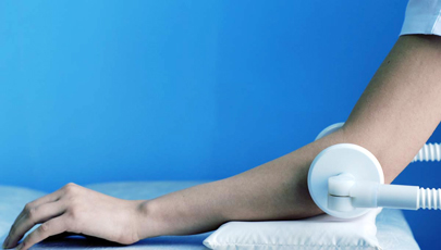
Physiotherapy modalities can aid in accelerating the reduction of swelling, inflammation, and soreness, as well as restoring the range of motion during the treatment of elbow bursitis.
Management of Purulent (Septic) Bursitis and Chronic Post-Traumatic Bursitis
- Purulent Bursitis: Patients with septic bursitis are often referred to a surgeon or may require hospitalization. Treatment includes:
- Antibiotics: Systemic antibiotics are essential, initially often broad-spectrum and then tailored based on culture and sensitivity results from aspirated bursal fluid.
- Aspiration or Incision and Drainage: A puncture (aspiration) of the bursa is often performed to obtain fluid for diagnosis and to relieve pressure. In many cases, surgical incision and drainage of the synovial bursa are necessary to adequately remove pus and infected material. The wound is then treated according to general principles of purulent surgery, which may involve packing or placement of a drain.
- Chronic Post-Traumatic Bursitis: This form, especially if there is blood (hemobursa) in the cavity of the synovial bursa (which provides a favorable environment for infection development), is often treated with surgical intervention (bursectomy - removal of the bursa). Relapses of the disease are also possible with chronic post-traumatic bursitis if the underlying irritation or trauma persists or if the bursa wall remains significantly thickened and inflamed.
Surgical removal of the bursa (bursectomy) may be considered for chronic, recurrent, or persistently painful bursitis that does not respond to conservative measures, or for septic bursitis requiring extensive debridement.
Differential Diagnosis of Joint Area Pain and Swelling
Pain and swelling around a joint can be caused by various conditions. It's important to differentiate bursitis from:
| Condition | Key Differentiating Features |
|---|---|
| Bursitis | Localized pain, tenderness, and swelling over a bursa. Pain often worse with specific movements that compress/stretch the bursa. Joint range of motion may be painful at extremes but often preserved passively if not directly compressing bursa. |
| Arthritis (e.g., Osteoarthritis, Rheumatoid Arthritis, Gout) | Pain, stiffness, swelling, and often restricted range of motion *within the joint itself*. Morning stiffness common in inflammatory arthritis. X-rays may show joint space narrowing, osteophytes, or erosions. Gout often has acute, severe attacks with redness and warmth over joint. |
| Tendinitis/Tendinopathy | Pain localized to a tendon, often worse with active movement or resisted contraction of the involved muscle. Tenderness on palpation of the tendon. Swelling may be present along the tendon sheath (tenosynovitis). |
| Ligament Sprain/Tear | History of acute injury/trauma. Localized pain, swelling, bruising over the ligament. Instability or pain with stress testing of the ligament. |
| Muscle Strain | Pain and tenderness within a muscle belly, often after overuse or sudden exertion. Pain with stretching or contracting the muscle. |
| Cellulitis | Spreading redness, warmth, swelling, and tenderness of the skin and subcutaneous tissue. Often associated with a break in the skin. Fever and systemic symptoms may be present. Borders usually indistinct. |
| Septic Arthritis | Infection within the joint. Severe pain, swelling, redness, warmth of the joint, significantly restricted range of motion (both active and passive), fever. A medical emergency requiring urgent joint aspiration and antibiotics. |
| Fracture | History of trauma. Severe pain, swelling, bruising, deformity, inability to bear weight or use the limb. Bony tenderness. X-ray confirms. |
| Tumor (Benign or Malignant) | Persistent, often progressive pain and/or swelling. May have night pain. Requires imaging (X-ray, CT, MRI) and possibly biopsy. |
Potential Complications of Bursitis
While often self-limiting or responsive to treatment, bursitis can lead to complications:
- Chronic Pain and Impaired Function: Persistent inflammation can lead to ongoing pain and difficulty using the affected joint/limb.
- Muscle Atrophy: Disuse due to pain can cause weakening and atrophy of surrounding muscles.
- Adhesive Capsulitis (Frozen Shoulder): Prolonged immobility of the shoulder due to bursitis can contribute to the development of a frozen shoulder.
- Calcific Bursitis: Deposition of calcium crystals within the bursa can cause chronic inflammation and pain.
- Rupture of the Bursa: Rare, can cause sudden worsening of pain and swelling.
- Spread of Infection (from Septic Bursitis): Infection can spread to adjacent soft tissues (cellulitis), bone (osteomyelitis), or into the bloodstream (sepsis), though this is uncommon with prompt treatment.
- Recurrence: Bursitis can recur, especially if the underlying cause (e.g., repetitive strain, biomechanical issues) is not addressed.
- Thickening of Bursa Wall (Chronic Bursitis): Can lead to persistent symptoms.
Prevention Strategies
Preventing bursitis often involves addressing risk factors:
- Avoiding Repetitive Motions or Prolonged Pressure: If your job or hobby involves repetitive tasks or pressure on certain joints, take regular breaks, vary activities, and use ergonomic aids or padding (e.g., knee pads for kneeling, elbow pads).
- Proper Technique and Warm-up: Use proper form during physical activities and sports. Warm up adequately before exercise and cool down afterward.
- Gradual Increase in Activity: Avoid sudden increases in the intensity or duration of exercise or physical activity.
- Maintaining a Healthy Weight: Reduces stress on joints.
- Stretching and Strengthening: Regular exercise to maintain flexibility and strength of muscles around joints can help improve biomechanics.
- Using Protective Gear: For activities that put stress on specific joints.
- Treating Underlying Conditions: Managing conditions like gout or rheumatoid arthritis can help prevent associated bursitis.
When to Seek Medical Attention
Consult a healthcare professional if you experience:
- Significant joint area pain or swelling that doesn't improve with home care (rest, ice) after a few days.
- Pain that is severe or rapidly worsening.
- Inability to move a joint.
- Signs of infection: increasing redness, warmth, tenderness, fever, or pus draining from the area.
- Recurrent episodes of bursitis.
- Pain that interferes with daily activities or sleep.
Early diagnosis and appropriate treatment can lead to faster recovery and prevent chronic problems.
References
- Aaron DL, Patel A, Kayiaros S, Calfee R. Four common types of bursitis: diagnosis and management. J Am Acad Orthop Surg. 2011 Jun;19(6):359-67.
- Williams CH, Park C, Co A. Bursitis. In: StatPearls [Internet]. Treasure Island (FL): StatPearls Publishing; 2023 Jan-.
- Fayad F, De Corla-Souza A, Le Bozec A, et al. Bursitis: when and how to treat it? Joint Bone Spine. 2003 May;70(3):170-6.
- Zimmerman B, Mikolich DJ, Lally EV. Septic bursitis. Semin Arthritis Rheum. 1995 Jun;24(6):391-410.
- Canoso JJ. Bursae, tendons, and ligaments. Clin Rheum Dis. 1981 Aug;7(2):189-221.
- Shbeeb MI, O'Duffy JD, Michet CJ Jr, Matteson EL. Evaluation of glucocorticosteroid injection for the treatment of olecranon bursitis. J Rheumatol. 1996 Dec;23(12):2104-6.
- Reilly D, Klinkhoff A. Bursitis. Can Fam Physician. 1990 Oct;36:1701-3.
See also
- Abscess
- Breast diseases (mastopathy, cyst, calcifications, fibroadenoma, intraductal papilloma, cancer)
- Bursitis
- Furuncle (boil)
- Ganglion cyst
- Hidradenitis suppurativa (HS)
- Ingrown toenail
- Lipoma (fatty tumor)
- Lymphostasis
- Paronychia, panaritium (whitlow or felon)
- Sebaceous cyst (epidermoid cyst)
- Tenosynovitis (infectious, stenosing)


