Understanding how muscles contract
Getting a grip on the sliding filament
A muscle contracts when all the sarcomeres in all the myofibrils in all the fibers (cells) contract all together. The sliding filament model describes the fine points of how this happens.
The key to the sliding filament model is the distinctive shapes of the protein molecules myosin and actin, and their partial overlap in the sarcomere. The special chemistry of ATP supplies the energy for the filaments’ movement. The following sections explain how sarcomeres create muscle contraction.
![]() The sarcomere is the functional unit within the myofibril. Sarcomeres line up end to end along the myofibril.
The sarcomere is the functional unit within the myofibril. Sarcomeres line up end to end along the myofibril.
Assembling a sarcomere
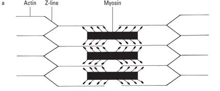
The sarcomere is composed of thick filaments and thin filaments. The thick filaments are molecules of the protein myosin, which is dense and rubbery. The thin filaments are made up of two strands of the lighter (less dense) protein actin, which is springy. The thin and thick filaments line up together in an orderly way to form a sarcomere. One end of a thin filament touches and adjoins the end of another thin filament forming the Z line that runs perpendicular to the filament axis. The sarcomere begins at one Z line and ends at the next Z line. The thick filaments line up precisely between the thin filaments. Sarcomeres and Z lines are shown in Figures 6-2 and 6-3.
The two types of filaments overlap only partially when the sarcomere is at rest. The partial overlap gives skeletal and cardiac muscle cells their striations: where thick and thin filaments overlap, the tissue appears dark (dark band); where only thin filaments are present, the tissue appears lighter (light band).
The myosin filaments have numerous club-shaped heads that point away from its center (toward the two Z lines). These heads rest nearly touching the myosin binding sites on the actin. Why, then, do they not link up? Wrapping around the actin is the troponin-tropomyosin complex. When a muscle is at rest, this protein covers up the binding sites. Until the myosin heads can link up with the actin, the fiber cannot contract. (See Figure 6-3a for a sarcomere at rest.)
Telling the fiber to contract
Motor neurons provide the stimulus for a muscle fiber to contract. From the end of its axon, a motor neuron releases acetylcholine (ACh), which binds to the muscle cell in a designated area called the motor end plate. This triggers the fiber to generate an impulse, which spreads through the sarcolemma (for more on nervous communication and impulse, see Chapter 7). The impulse is spread deep into the cell by tunnels called T-tubules. When the impulse reaches a sarcoplasmic reticulum (SR), it is triggered to release the calcium ions stored there. The ions move to the sarcomeres, allowing contraction to begin. Triggering relaxation is a passive process. The neuron simply stops releasing ACh. Muscle impulse stops and the calcium ions return to the SR.
Contracting and releasing the sarcomere
After calcium is released from the SR, it attaches to the troponin. In doing so, it is pulled away from the actin — like pulling a push pin out of a bulletin board. Because troponin is still tightly bound to the tropomyosin, the whole protein complex slides around the actin, exposing the binding sites. Now the myosin heads can link up with the actin forming cross bridges, all without the use of energy. However, to actually cause a contraction, we must shorten the sarcomere.
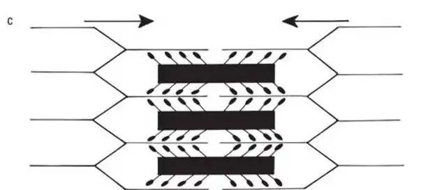
When the cross bridges form, the myosin heads immediately bend, pulling the ends of the sarcomere (Z lines) closer together. ATP will then bind to the myosin, causing it to let go of the actin. The energy in ATP causes the heads to cock back into their original position. They will then form a new cross bridge except it is now further down the actin (similar to how a ratchet wrench works). These power strokes continue — grab, pull in, let go, cock back — as long as the binding sites are uncovered and ATP is present. Refer to Figure 6-3 to get a picture of this process.
The sarcomere shortens as the filaments slide past each other. Because the myofibrils are made of sarcomeres that share Z lines (refer to Figure 6-2), and this shortening is occurring in all of them, the two ends of the myofibril are pulled noticeably closer together. Because a muscle fiber is packed full of myofibrils, it is shortened as its ends are pulled together, too. A skeletal muscle is made of numerous fibers bundled in parallel to each other so its ends are pulled together as well. The muscle contracts, pulling whatever it’s attached to closer together, all with the force generated by the overlapping of these microscopic filaments (but multiplied thousands of times over).
![]() The filaments (actin and myosin) do not shrink. They maintain their length at all times. The myosin filaments also do not move. Actin is pulled toward the center, progressively increasing the amount of overlap with the myosin (refer to Figure 6-3c).
The filaments (actin and myosin) do not shrink. They maintain their length at all times. The myosin filaments also do not move. Actin is pulled toward the center, progressively increasing the amount of overlap with the myosin (refer to Figure 6-3c).
In order to relax, the fiber simply stops being told to contract (the motor neuron halts its stimulation). This causes the SR to call back the calcium ions like a mother calling her kids back home for dinner. They happily oblige, letting go of the troponin, which will pin back into the actin. This slides the tropomyosin over the top of the binding sites again, knocking off any of the myosin heads that were attached (breaking the cross bridges). The actin will then slowly slide back restoring the length of the sarcomere to its original position.
The last contraction
The time comes for every animal — humans included — to die. The fact that every animal gets cold and stiff tells others when that time has come. Do you know why? The cells no longer make ATP.
At the moment of death, the lungs stop filling with oxygen, the heart stops pumping blood through the body, and the brain stops sending signals. The cells — without incoming oxygen, nutrients, or stimulus from the brain — cease performing their metabolic reactions. So ATP can no longer be produced.
Without ATP flooding the myofibrils, contractions can’t occur, but neither can the last step of muscle contraction, the step that allows the muscles to relax. In order for a myofibril to relax, ATP must hook onto myosin and dissolve the actin-myosin cross-bridges. But when ATP is unavailable to generate a subsequent contraction, the last contraction becomes permanent, and the corpse stiffens. Rigor mortis, which means rigidity of death, occurs in every muscle throughout the body. And remember that movement of muscles generates heat, so when the muscles stop their physiological reactions and warm blood stops flowing through the blood vessels, the corpse gets cold.
See also
- Locating Physiology on the Web of Knowledge
- Chapter 1. Anatomy and Physiology: The Big Picture
- Chapter 2. What Your Body Does All Day
- Chapter 3. A Bit about Cell Biology
- Sizing Up the Structural Layers
- Chapter 4. Getting the Skinny on Skin, Hair, and Nails
- Chapter 5. Scrutinizing the Skeletal System
- Chapter 6. Muscles: Setting You in Motion
- Talking to Yourself
- Chapter 7. The Nervous System: Your Body’s Circuit Board
- Chapter 8. The Endocrine System: Releasing Chemical Messages
- Exploring the Inner Workings of the Body
- Chapter 9. The Cardiovascular System: Getting Your Blood Pumping
- Chapter 10. The Respiratory System: Breathing Life into Your Body
- Chapter 11. The Digestive System: Beginning the Breakdown
- Chapter 12. The Urinary System: Cleaning Up the Act
- Chapter 13. The Lymphatic System: Living in a Microbe Jungle
- Life’s Rich Pageant: Reproduction and Development
- Chapter 14. The Reproductive System
- Chapter 15. Change and Development over the Life Span
- The Part of Tens
- Chapter 16. Ten (Or So) Chemistry Concepts Related to Anatomy and Physiology
- Chapter 17. Ten Phabulous Physiology Phacts
- Supplemental Images


