Taking a tour of the skeletal muscles
Naming the skeletal muscles
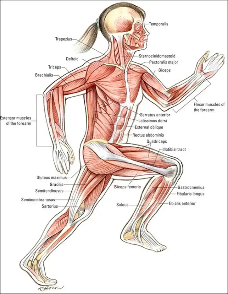
Get ready, because we’re about to tell you the muscle names from head to toe — literally. Check out the "Muscular System" color plate in the center of the book as you go through this section.
To name muscles, anatomists had to come up with a set of rules to follow so the names would make sense. They chose to focus on certain characteristics to derive each muscle’s Latin name. As you go through the following sections, refer to Chapter 1 if necessary for the names of the body regions. Examples of characteristics in muscle names are given in Table 6-2.
TABLE 6-2 Characteristics in Muscle Names
| Characteristics | Examples |
| Muscle size | The largest muscle in the buttocks is the gluteus maximus (maximus means large in Latin); a smaller muscle in the buttocks is the gluteus minimus (minimus means small in Latin). |
| Muscle location | The frontalis muscle lies on top of the skull’s frontal bone. |
| Muscle shape | The deltoid muscle, shaped like a triangle, comes from delta — the Greek alphabet’s fourth letter, which is also shaped like a triangle. |
| Muscle action | The extensor digitorum is a muscle that extends the fingers or digits. |
| Number of muscle attachments | The biceps brachii attaches to bone in two locations, whereas the triceps brachii attaches to bone in three locations. |
| Muscle fiber direction | The rectus abdominis muscle runs vertically along your abdomen (rectus means straight in Latin). |
Starting at the top
Your head contains muscles that perform three basic functions: chewing, making facial expressions, and moving your neck. Ear wiggling falls into this category, too.
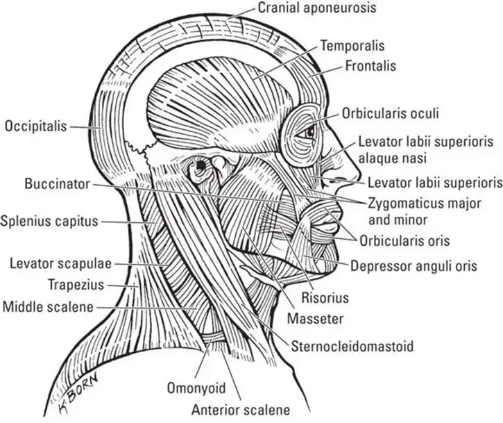
To chew, you use the muscles of mastication (a big, fancy word that means "chewing"). The masseter, a muscle that runs from the zygomatic bone (your cheekbone) to the mandible (your lower jaw), is the prime mover for mastication, so its name is based on its action (masseter, mastication). The fan-shaped temporalis muscle works with the masseter to allow you to close your jaw. It lies on top of the skull’s temporal bone, so its name is based on its location. Figure 6-4 shows the muscles of the head and neck.
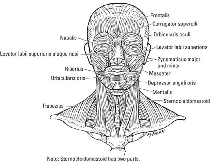
To smile, frown, or make a funny face, you use several muscles. The frontalis muscle along with a tiny muscle called the corrugator supercilii raises your eyebrows and gives you a worried or angry look when wrinkling your brow. (Think of the appearance of corrugated cardboard, and then feel the skin between your eyebrows when you wrinkle your brow.) The orbicularis oculi muscle surrounds the eye (the word orbit, as in orbicularis, means "to encircle"; oculi refers to the eye). This muscle allows you to blink your eyes and close your eyelids, but it also gives you those little crow’s feet at the corners of your eyes. The orbicularis oris surrounds the mouth. (Or refers to mouth, as in "oral.") You use this muscle to pucker up for a kiss. Figure 6-5 shows the facial muscles.
If you play the trumpet or another instrument that requires you to blow out, you’re well aware of what your buccinator muscle does. This muscle is in your cheek. (Bucc means "cheek," as in the word buccal, which refers to the cheek area.) It allows you to whistle and also helps keep food in contact with your teeth as you chew. Remember that your zygomatic is your cheekbone? Well, the zygomaticus muscle is a branched muscle that runs from your cheekbone to the corners of your mouth. This muscle pulls your mouth up into a smile when the mood strikes you.
When you want to nod yes, no, or tilt your head into a maybe so, your neck muscles come into play. You have two sternocleidomastoid muscles, one on each side of your neck. We know this is a long name, but the name reflects the locations of its attachments: the sternum, clavicle, and mastoid process of the skull’s temporal bone. When both sternocleidomastoid muscles contract, you can bring your head down toward your chest and flex your neck. When you turn your head to the side, one sternocleidomastoid muscle contracts — the one on the opposite side of the direction your head is turned. So if you turn your head to the left, your right sternocleidomastoid muscle contracts, and vice versa. If you lean your head back to look up at the sky or to shrug your shoulders, your trapezius muscle allows you to do so.
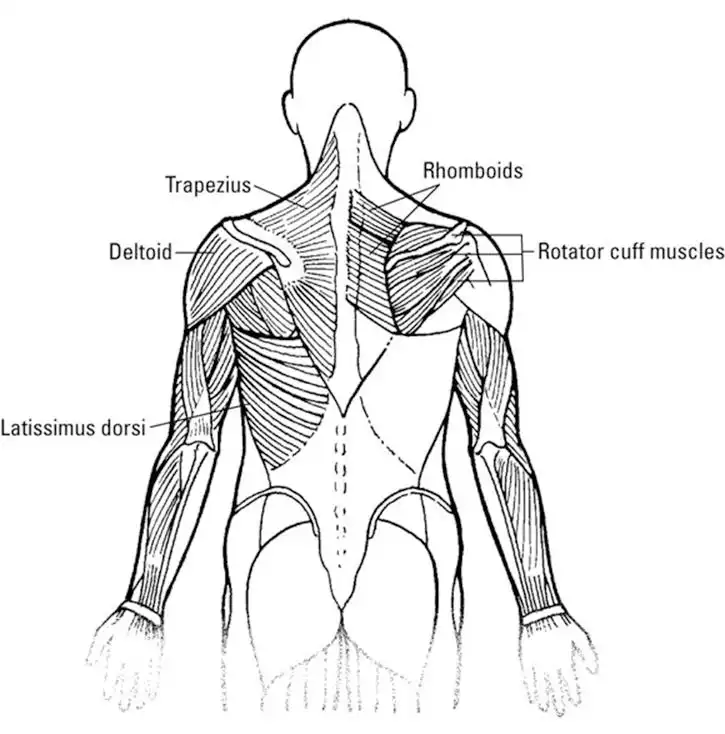
The trapezius is an antagonist to the sternocleidomastoid muscle. If you remember basic geometry, you can see that the trapezius is shaped like a trapezoid. It runs from the base of your skull to your thoracic vertebrae and connects to your scapula (shoulder blades). Therefore, the trapezius and sternocleidomastoid muscles connect your head to your torso and provide a nice segue to the next section. The trapezius is shown in Figure 6-7.
Twisting the torso
![]() The torso muscles have important functions. They not only give support to your body but also connect to your limbs to allow movement, allow you to inhale and exhale, and protect your internal organs. In this section, we cover the muscles that run along the front of you (called your anterior or ventral side) and then cover the muscles of your back (your posterior or dorsal side).
The torso muscles have important functions. They not only give support to your body but also connect to your limbs to allow movement, allow you to inhale and exhale, and protect your internal organs. In this section, we cover the muscles that run along the front of you (called your anterior or ventral side) and then cover the muscles of your back (your posterior or dorsal side).
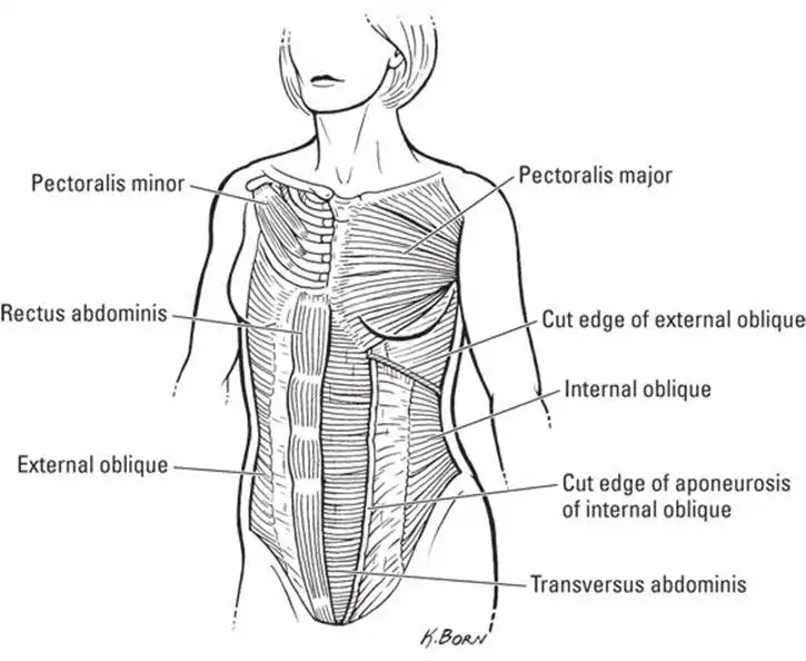
In your chest (see Figure 6-6), your pectoralis major muscles connect your torso at the sternum and collarbones (clavicles) to your upper limbs at the humerus bone in the upper arm. Your "pecs" also help to protect your ribs, heart, and lungs. You can feel your pectoralis major muscle working when you move your arm across your chest. Also in your chest are the muscles between and around the ribs. The internal intercostal muscles help to raise and lower your rib cage as you breathe. However, the torso’s largest muscles are the abdominal muscles.
![]() The abdominal muscles really form the center of your body. If the abdominal muscles are weak, the back is weak because the abdominal muscles help to flex the vertebral column. So if the vertebral column doesn’t flex easily, the muscles attached to it can become strained and weak. And the muscles of the abdomen and back join to the upper and lower limbs. Therefore, if the abdomen and back are weak, the limbs can have problems.
The abdominal muscles really form the center of your body. If the abdominal muscles are weak, the back is weak because the abdominal muscles help to flex the vertebral column. So if the vertebral column doesn’t flex easily, the muscles attached to it can become strained and weak. And the muscles of the abdomen and back join to the upper and lower limbs. Therefore, if the abdomen and back are weak, the limbs can have problems.
The muscles of the abdomen are thin, but the fact that these muscle fibers run in different directions increases their strength. This woven effect makes the tissues much stronger than they would be if they all went in the same direction. Think about how a child connects building blocks. Laying a top layer of blocks perpendicular to the blocks underneath helps the structure stay together, which is similar to how the abdominal muscle tissues provide strength and stability.
The "six-pack" muscle of the abdomen, the rectus abdominis, forms the front layer of the abdominal muscles, and it runs from the pubic bone up to the ribs and sternum. The function of the rectus abdominis muscle is to hold in the organs of the abdomino-pelvic cavity and allow the vertebral column to flex.
Other layers of abdominal muscles also help to hold in your organs on the side of your abdomen and provide strength to your body’s core. The external oblique muscles attach to the eight lower ribs and run downward toward the middle of your body (slanting toward the pelvis). The internal oblique muscles lie underneath the external oblique (makes sense, eh?) at right angles to the external oblique muscles. The internal oblique muscles extend from the top of the hip at the iliac crest to the lower ribs.
Together, the external and internal oblique muscles form an X, essentially strapping together the abdomen. The abdomen’s deepest muscle, the transversus abdominis, runs horizontally across the abdomen; its function is to tighten the abdominal wall, push the diaphragm upward to help with breathing, and help the body bend forward. The transversus abdominis is connected to the lower ribs and lumbar vertebrae and wraps around to the pubic crest and linea alba.The linea alba ("white line") is a band of connective tissue that runs vertically down the front of the abdomen from the xiphoid process at the bottom of the sternum to the pubic symphysis (the strip of connective tissue that joins the hip bones).
The muscles in your back (refer to Figure 6-7) serve to provide strength, join your torso to your upper and lower limbs, and protect organs that lie toward the back of your trunk (such as your kidneys). The deltoid muscle joins the shoulder to the collarbone, scapula, and humerus. This muscle is shaped like a triangle (think of the Greek letter delta: Δ). The deltoid muscle helps you raise your arm up to the side (that is, laterally). The latissimus dorsi muscleis a wide muscle that’s also shaped like a triangle. It originates at the lower part of the spine (thoracic and lumbar vertebrae) and runs upward on a slant to the humerus. Your "lats" allow you to move your arm down if you have it raised, and also to reach, such as when you’re climbing or swimming.
Spreading your wings
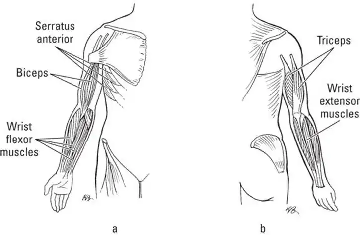
Your upper limbs have a wide range of motions. Obviously, your upper limbs are connected to your torso. One of the muscles that provides that connection, the serratus anterior, is below your armpit (the anatomic term for armpit is axilla) and on the side of your chest. The serratus anterior muscle connects to the scapula and the upper ribs. You use this muscle when you push something or raise your arm higher than horizontal. Its action pulls the scapula downward and forward.
Although the biceps brachii and triceps brachii are muscles located in the top (anterior) part of your upper arm, their actions allow your forearm (lower arm) to move. Figure 6-8a shows an anterior view of the upper limb. The name biceps refers to this muscle’s two origins (points of attachment); it attaches to the scapula in two places. From there, it runs to the radius of the forearm (its point of insertion). The triceps brachii is the only muscle that runs along the back (posterior) side of the upper arm. Figure 6-8b shows a posterior view of the upper limb. The name triceps refers to the fact that it has three attachments: one on the scapula and two on the humerus. It runs to the ulna of the forearm. You can feel this muscle in motion when you push or punch. Other muscles of the arm include the brachioradialis, which helps you flex your arm at the elbow, and the supinator, which rotates your arm from a palm-down position to a palm-up position (remember the word supinator has the word "up" in it).
Your forearm contains muscles that control the fine movements of your fingers. When you type or play the piano, you’re using your extensor digitorum and flexor digitorum muscles to raise and lower your fingers onto the keyboard and move them to the different rows of keys. As you lift your hands off the keyboard, the muscles of your wrist kick into gear. The flexor carpi radialis (attached to the radius bone) and flexor carpi ulnaris (attached to the ulna bone) allow your wrist to flex forward or downward. The extensor carpi radialis longus (which passes by the carpal bones), the extensor carpi radialis brevis, and the extensor carpi ulnaris allow your wrist to extend; that is, bend upward.
![]() The muscles in your upper arm move your forearm. The muscles of your forearm move your wrists, hands, and fingers. There are no muscles in your fingers — only the tendons that connect to those bones.
The muscles in your upper arm move your forearm. The muscles of your forearm move your wrists, hands, and fingers. There are no muscles in your fingers — only the tendons that connect to those bones.
Thumbs up!
A key feature of all primates is a prehensile thumb; that is, a thumb that’s adapted for grasping objects. Many animals have digitlike structures, but only primates can grasp things with their hands. And the only way to grasp things is to have a thumb.
Imagine having webbing between your four fingers so that you couldn’t spread them apart; you wouldn’t be able to pick things up. That’s why animals such as dogs, cats, and birds hold things in their mouth (or beak). But primates — apes, monkeys, and humans — can easily grasp things between their thumb and fingers. However, of those primates, only humans have an opposable thumb (one that can touch each of the other fingers; the thumb can be "opposite" from each finger).
Because of the ability to oppose the thumb to each finger, the muscles in your digits are capable of performing minute movements. As you touch your thumb to your pinky finger, your palm becomes arched, which only happens in humans because of short bones in the pinky and an opposable thumb.
Getting a leg up
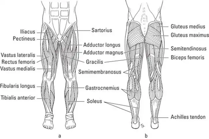
Your lower limbs are connected to your buttocks, and your buttocks are connected to your hips. The iliopsoas connects your lower limb to your torso and consists of two smaller muscles: the psoas major, which joins the thigh to the vertebral column, and the iliacus, which joins the hipbone’s ilius to the thigh’s femur bone. Originating on the iliac spine of the hip and joining to the inside surface of the tibia (a bone in your shin), the sartorius muscle is a long, thin muscle that runs from the hip to the inside of the knee (see Figure 6-9a). These muscles stabilize your lower limbs and provide strength for them to support your body’s weight and to balance your body against the pressure of gravity.
Some muscles in the lower limb allow the thigh to move in a variety of positions. The buttocks muscles allow you to straighten your lower limb at the hip and extend your thigh when you walk, climb, or jump. The gluteus maximus — the largest muscle in the buttocks — is the largest muscle in the body (see Figure 6-9b). The gluteus maximus is antagonistic to the stabilizing iliopsoas muscle, which flexes your thigh. The gluteus medius muscle,which lies behind the gluteus maximus, allows you to raise your leg to the side so that you can form a 90° angle with your two legs (this action is abduction of the thigh). Several muscles serve as adductors; that is, they move an abducted thigh back toward the midline. These muscles include the pectineus and adductor longus, which become injured when you "pull a groin muscle," as well as the adductor magnus and gracilis, which run along the inside of your thigh.
Other muscles in the thigh serve to move the lower leg. Along your thigh’s front and lateral side, four muscles work together to allow you to kick. These four muscles — the rectus femoris, vastus lateralis, vastus medialis, and vastus intermedius — are better known as the quadriceps (quadriceps femoris). Quad means "four", as in quadrilateral or quadrant. Refer to Figure 6-9a.
The hamstrings are a group of muscles that are antagonistic to the quadriceps. The hamstrings — the biceps femoris, semimembranosus, and semitendinosus — run down the back of the thigh (refer to Figure 6-9b) and allow you to flex your lower leg and extend your hip. They originate on the ischium of the hipbone and join (insert at) to the tibia of the lower leg. You can feel the tendons of your hamstring muscles behind your knee.
Your lower leg’s shin and calf muscles move your ankle and foot. The gastrocnemius, better known as the "calf muscle", begins (originates) at the femur (thigh bone) and joins (inserts at) the Achilles tendon that runs behind your heel. You can feel your gastrocnemius muscle contracting when you stand on your toes. The antagonist of the gastrocnemius, the tibialis anterior, starts on the surface of the tibia (shinbone), runs along the shin, and connects to your ankle’s metatarsal bones. You can feel this muscle contract when you raise your toes and keep your heel on the floor. The fibular longus and fibular brevis (brevis meaning "short", as in brevity) run along the outside of the lower leg and join the fibula to the ankle bones. In doing so, the fibular muscles help to move the foot. The extensor digitorum longus and the flexor digitorum longus muscles join the tibia to the feet and allow you to extend and flex your toes, respectively, like your fingers.
Where did these names come from?
Some muscle names have a pretty interesting history. Take the hamstring muscles and the sartorius muscle. First the hamstrings — ham may make you think of pigs, and yes, pigs have hamstrings in their legs. And the biceps femoris, semimembranosus, and semitendinosus muscles have the same strong tendons in a pig as they do in you. When butchers smoked hams (thigh meat from a pig), they hung the hams on hooks in the smokehouse by these ropelike tendons, which generated the name "hamstrings". Nobody said butchers were creative.
The sartorius muscle goes into action when you sit cross-legged, like tailors used to do when they pinned hems or cuffs (and maybe still do). So the sartorius muscle is sometimes referred to as the "tailor’s muscle." And guess what means "tailor" in Latin? Yep, sartor.
See also
- Locating Physiology on the Web of Knowledge
- Chapter 1. Anatomy and Physiology: The Big Picture
- Chapter 2. What Your Body Does All Day
- Chapter 3. A Bit about Cell Biology
- Sizing Up the Structural Layers
- Chapter 4. Getting the Skinny on Skin, Hair, and Nails
- Chapter 5. Scrutinizing the Skeletal System
- Chapter 6. Muscles: Setting You in Motion
- Talking to Yourself
- Chapter 7. The Nervous System: Your Body’s Circuit Board
- Chapter 8. The Endocrine System: Releasing Chemical Messages
- Exploring the Inner Workings of the Body
- Chapter 9. The Cardiovascular System: Getting Your Blood Pumping
- Chapter 10. The Respiratory System: Breathing Life into Your Body
- Chapter 11. The Digestive System: Beginning the Breakdown
- Chapter 12. The Urinary System: Cleaning Up the Act
- Chapter 13. The Lymphatic System: Living in a Microbe Jungle
- Life’s Rich Pageant: Reproduction and Development
- Chapter 14. The Reproductive System
- Chapter 15. Change and Development over the Life Span
- The Part of Tens
- Chapter 16. Ten (Or So) Chemistry Concepts Related to Anatomy and Physiology
- Chapter 17. Ten Phabulous Physiology Phacts
- Supplemental Images

