Delineating life’s levels of organization
Organizing yourself on many levels
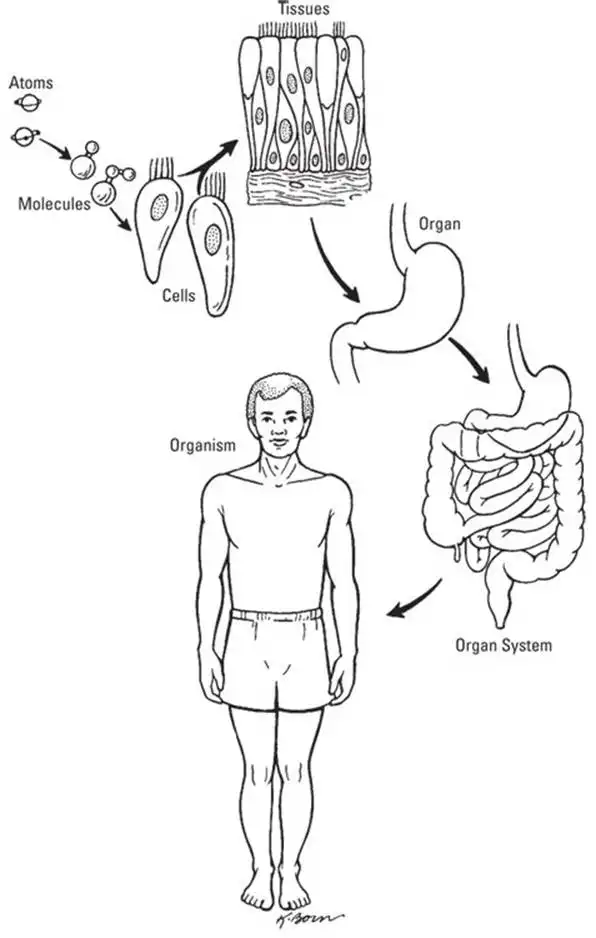
Anatomy and physiology are concerned with the level of the individual body, what scientists call the organism. However, you can’t merely focus on the whole and ignore the role of the parts. The life processes of the organism are built and maintained at several physical levels, which biologists call levels of organization: the cellular level, the tissue level, the organ level, the organ system level, and the organism level (see Figure 1-5). In this section, we review these levels, starting at the bottom.
Level I: The cellular level
If you examine a sample of any human tissue under a microscope, you see cells, possibly millions of them. All living things are made of cells. In fact, "having a cellular level of organization" is inherent in any definition of "organism". The work of the body actually occurs in the cells; for example, your whole heart beats to push blood around your body because of what happens inside the cells that create its walls.
Level II: The tissue level
A tissue is a structure made of many cells — usually several different kinds of cells — that performs a specific function. Tissues are divided into four categories:
- Connective tissue serves to support body parts and bind them together. Tissues as different as bone and blood are classified as connective tissue.
- Epithelial tissue (epithelium) functions to line and cover organs as well as carry out absorption and secretion. The outer layer of the skin is made up of epithelial tissue.
- Muscle tissue — surprise! — is found in the muscles, which allow your body parts to move; in the walls of hollow organs (such as intestines and blood vessels) to help move their contents along; and in the heart to move blood along via the acts of contraction and relaxation. (Find out more about muscles in Chapter 6)
- Nervous tissue transmits impulses and forms nerves. Brain tissue is nervous tissue. (We talk about the nervous system in Chapter 7)
Level III: The organ level
An organ is a group of tissues assembled to perform a specialized physiological function. For example, the stomach is an organ that has the specific physiological function of breaking down food. By definition, an organ is made up of at least two different tissue types; many organs contain tissues of all four types. Although we can name and describe all four tissue types that make up all organs, as we do in the preceding section, listing all the organs in the body wouldn’t be so easy.
Level IV: The organ system level
Human anatomists and physiologists have divided the human body into organ systems, groups of organs that work together to meet a major physiological need. For example, the digestive system is one of the organ systems responsible for obtaining energy from the environment. Realize, though, that this is not a classification system for your organs. The organs that "belong" to one system can have functions integral to another system. The pancreas, for example, produces enzymes vital to the breakdown of our food (digestion), as well as hormones for the maintenance of our homeostasis (endocrine).
The chapter structure of this book is based on the definition of organ systems.
Level V: The organism level
The whole enchilada. The real "you". As we study organ systems, organs, tissues, and cells, we’re always looking at how they support you on the organism level.
Taking pictures of your insides
For early anatomists like Hippocrates and da Vinci, the images they had were the sketches they made for themselves. The drawings made by Andreas Vesalius were compiled into the first anatomical atlas and the accuracy, considering it was the 16th century, is impressive. However, it is a German physicist named Wilhelm Conrad Roentgen who’s remembered as "the father of medical imaging". In 1895, Roentgen changed the game by recording the first image of the internal parts of a living human: an X-ray image of his wife’s hand. By 1900, X-rays were in widespread use for the early detection of tuberculosis, at that time a common cause of death. X-rays are beams of radiation emitted from a machine toward the patient’s body, and X-ray images show details only of hard tissues, like bone, that reflect the radiation. In this way, they’re similar to photographs. Refinements and enhancements of X-ray techniques were developed all through the 20th century, with extensive use and major advances during World War II. The X-ray is still a widely used method for medical diagnosis, not just for bone breaks but for screening for signs of disease, especially tumors.
In the 1970s, computer technology took off, taking medical imaging technology with it. Digital imaging techniques began to be applied to convert multiple flat-slice images into one three-dimensional image. The first technology of this sort was called computed axial tomography (commonly called a CAT or CT scan). The technique combines multiple X-ray images of varying depths into images of whole structures inside the body. Contrast dye can be used to highlight particular areas, which is especially useful for a quick assessment (for example, after a trauma).
Another class of imaging technology utilizing radiation is positron emission tomography (PET). A radioactive isotope can be attached to a specific molecule — a drug, for example. After administering the drug to the patient, the isotope emits radiation, which can be traced and followed with radiation detectors. This is especially useful for testing the efficacy of drugs in a clinical research setting. It’s unique in that the scan provides information of organ function on a cellular level.
Ultrasound imaging technology uses the echoes of sound waves sent into the body to generate a signal that a computer turns into a real-time image of anatomy and physiology. Ultrasound can also produce audible sounds, so the anatomist or physiologist can, for example, watch the pulsations of an artery while hearing the sound of the blood flowing through it. Although all these technologies are considered noninvasive, ultrasound is the least invasive of all (no radiation) so it’s used more freely, especially in sensitive situations like pregnancy.
Magnetic resonance imaging (MRI) utilizes magnetic fields and radio pulses to create an image of the interior. Soft tissue structures are more difficult to scan using other methods, especially those found underneath bone. The resulting 3D image can pinpoint anomalies within an organ, often in great detail. Since the early 1990s, neuroscientists have been using a type of specialized MRI scan, called functional MRI (fMRI), to acquire images of the brain. Images can be recorded over time, and the active areas of the brain "light up" on the scan, showing which parts are active during specific tasks. Basically, fMRI enables scientists to watch a patient’s or research subject’s thoughts as he or she is thinking them!
Digital imaging technologies produce images that are extremely clear and detailed. The images can be produced much more quickly and cheaply than older technologies allowed for, and the images can be easily duplicated, transmitted, and stored. The amount of anatomical and physiological knowledge that digital imaging technologies have helped generate over the past 30 years has transformed biological and medical science. As the techniques are continually researched and developed, our understanding of physiology and accuracy in diagnostics will continue to improve.
See also
- Locating Physiology on the Web of Knowledge
- Chapter 1. Anatomy and Physiology: The Big Picture
- Chapter 2. What Your Body Does All Day
- Chapter 3. A Bit about Cell Biology
- Sizing Up the Structural Layers
- Chapter 4. Getting the Skinny on Skin, Hair, and Nails
- Chapter 5. Scrutinizing the Skeletal System
- Chapter 6. Muscles: Setting You in Motion
- Talking to Yourself
- Chapter 7. The Nervous System: Your Body’s Circuit Board
- Chapter 8. The Endocrine System: Releasing Chemical Messages
- Exploring the Inner Workings of the Body
- Chapter 9. The Cardiovascular System: Getting Your Blood Pumping
- Chapter 10. The Respiratory System: Breathing Life into Your Body
- Chapter 11. The Digestive System: Beginning the Breakdown
- Chapter 12. The Urinary System: Cleaning Up the Act
- Chapter 13. The Lymphatic System: Living in a Microbe Jungle
- Life’s Rich Pageant: Reproduction and Development
- Chapter 14. The Reproductive System
- Chapter 15. Change and Development over the Life Span
- The Part of Tens
- Chapter 16. Ten (Or So) Chemistry Concepts Related to Anatomy and Physiology
- Chapter 17. Ten Phabulous Physiology Phacts
- Supplemental Images


