Looking separately at the axial and appendicular skeletons
- The axial skeleton
- Keeping your head up: the skull
- Cracking the cranium
- Facing the facial bones
- Sniffing out the sinuses
- The bone that floats
- Setting you straight on the curved spinal column
- Being caged can be a good thing
- The appendicular skeleton
- Wearing girdles: everybody has two
- Why weight-bearing exercise is good for you
- Why women have bigger hips than men
- Going out on a limb:arms and legs
- Giving you a hand with hands (and arms and elbows)
- Getting a leg up on your lower limbs
- Strong feet, firm foundation
The axial skeleton
The axial skeleton consists of the bones that lie along the midline (center) of your body, such as your vertebral column (backbone). An easy way to remember what bones make up the axial skeleton is to think of the vertebral column running down the middle of your body and then the bones that are directly attached to it — the thoracic cage (rib cage) and the skull.
The following sections give you a closer look at the main parts of the axial skeleton.
Keeping your head up: the skull
Rather than one big piece of bone, like a cap that fits over the brain, the skull comprises the cranial and the facial bones.
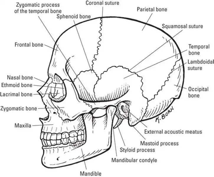
A human skull (see Figure 5-3) is made up of the cranium, which is formed of several bones, and the facial bones. The facial bones contain cavities called the sinuses, which do have a purpose other than harboring upper respiratory infections.
Cracking the cranium
The eight bones of your cranium protect your brain and have immovable joints between them called sutures. (These look a lot like the sutures, or stitches, that you may receive to close an incision or wound.) The bones of the cranium that are joined together by sutures include the following:
- Frontal bone: Gives shape to the forehead and part of the eye sockets.
- Parietal bones: Two bones that form the roof and sides of the cranium.
- Occipital bone: Forms the back of the skull and the base of the cranium. The foramen magnum, an opening in the occipital bone, allows the spinal cord to pass into the skull and join the brain.
- Temporal bones: Form the sides of the cranium near the temples. The temporal bone on each side of your head contains the following structures:
- External auditory meatus: The opening to your ear canal
- Mandibular fossa: Joins with the mandible (the lower jaw)
- Mastoid process: Provides a place for neck muscles to join your head
- Styloid process: Serves as an attachment site for muscles of the tongue and larynx (voice box)
- Ethmoid bone: Contains several sections, called plates. Forms the medial (inside) part of the eye sockets and much of the nasal cavity.
- Sphenoid bone: Shaped like a butterfly or a saddle (depending on how you look at it), the sphenoid forms the floor of the cranium and the back and lateral sides of the eye sockets (orbits). A central, sunken portion of the sphenoid bone called the sella turcica shelters the pituitary gland, which is very important in controlling major functions of the body. (See Chapter 8 for more on the pituitary gland.)
Facing the facial bones
The bones that form facial structures are:
- Lacrimal bones: Two tiny bones on the inside walls of the orbits. A groove between the lacrimal bones in the eye sockets and the nose forms the nasolacrimal canal. Tears flow across the eyeball and through that canal into your nasal cavity, which explains why your nose "runs" when you cry.
- Mandible: The lower jaw and the only movable bone of the skull.
- Maxillae: Two bones that form the upper jaw, part of the hard palate (roof of your mouth), and bottom of the orbits.
- Nasal bones: Two rectangular-shaped bones that form the bridge of your nose. The lower, movable portion of your nose is made of cartilage.
- Palatine bones: Form the back portion of the hard palate and is the floor of the nasal cavity.
- Vomer bone: Joins the ethmoid bone to form the nasal septum — that part of your nose that can be deviated by a strong left hook.
- Zygomatic bones: Form the cheekbones and the lateral (outer) sides of the orbits.
Sniffing out the sinuses
The sinuses allow air into the skull, making it much lighter. The air in your sinuses also gives resonance to your voice, which means that when you talk, the sound waves reverberate in your sinuses.
Several types of sinuses are named for their location:
- The frontal sinus is a hollowed-out area in the frontal bone.
- Mastoid sinuses drain into the middle ear.
- Maxillary sinuses are large and within the bones of the upper jaw (the maxilla).
- Paranasal sinuses consist of the frontal, sphenoidal, and ethmoidal sinuses, which, along with the maxillary sinuses, drain into the nose (para- means "near"; nas- means "nose").
The bone that floats
The hyoid bone, a tiny, U-shaped bone that resides just above your larynx (voice box), anchors the tongue and muscles used during swallowing. However, the hyoid bone itself isn’t attached to anything. It’s the only bone in the body that does not articulate (connect) with another bone. The hyoid bone hangs by ligaments attached to the styloid processes of the temporal bones.
Setting you straight on the curved spinal column
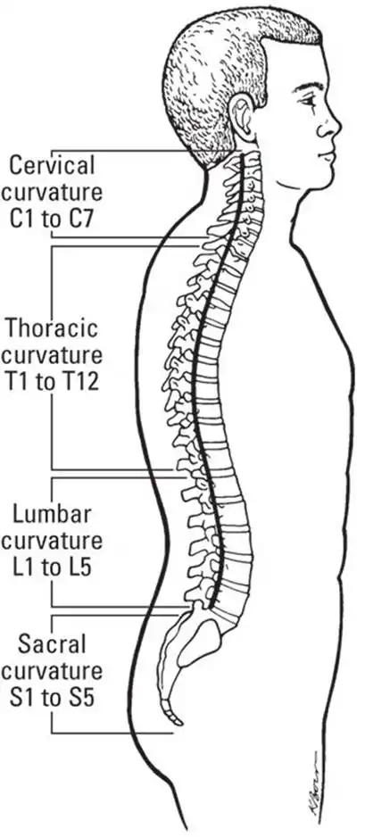
The spinal column (see Figure 5-4) begins within the skull and extends down to the pelvis. It’s made up of 33 bones in all: 24 separate bones called vertebrae (singular, vertebra), plus the fused bones of the sacrum and the coccyx. Between each vertebra is an intervertebral disc made of fibrocartilage for shock absorption.
Your spinal column is the central support for the upper body, carrying most of the weight of your head, chest, and arms. Together with the muscles and ligaments of your back, your spinal column enables you to walk upright.
![]() An important purpose of the vertebral column is to protect your spinal cord, the big data pipe between your body and your brain. Nearly all your nerves are connected, either directly or through networked branches, to the spinal cord, which runs directly into the brain through the opening in the skull called the foramen magnum. Turn to Chapter 7 for more information about the spinal cord.
An important purpose of the vertebral column is to protect your spinal cord, the big data pipe between your body and your brain. Nearly all your nerves are connected, either directly or through networked branches, to the spinal cord, which runs directly into the brain through the opening in the skull called the foramen magnum. Turn to Chapter 7 for more information about the spinal cord.
If you look at the spine from the side, you notice that it curves four times: inward, outward, inward, and outward. The curvature of the spine helps it absorb shock and pressure much better than if the spine were straight. A curved spine also affords more balance by better distributing the weight of the skull over the pelvic bones, which is needed to walk upright. A curved spine keeps you from being top-heavy. Each curvature spans a region of the spine: cervical, thoracic, lumbar, and sacral. The number of vertebrae in each region and some important vertebral features are given in Table 5-2.
TABLE 5-2 Regions of the Vertebral Column
| Region | Number of Vertebrae | Features |
| Cervical | 7 | The skull attaches at the top of this region to the vertebrae called the atlas. |
| Thoracic | 12 | The ribs attach to this region. |
| Lumbar | 5 | Commonly referred to as the small of the back, it takes the most stress. |
| Sacral | 5 (fused into one; the sacrum) | The sacrum forms a joint with the hipbones and the last lumbar vertebra. |
| Coccygeal | 4 (fused into one; the coccyx, also called the tailbone) | The coccyx absorbs the shock of sitting. |
The vertebral column also provides places for other bones to attach. The skull is attached to the top of the cervical spine. The first cervical vertebra (abbreviated C-1; "C" for cervical, "1" for first) is the atlas, which supports the head and allows it to move forward and back (for example, the "yes" movement). The second cervical vertebra (C-2) is called the axis, and it allows the head to pivot and turn side to side (that is, the "no" movement).
![]() You can differentiate these two important bones by recalling the Greek story about Atlas, who held the world on his shoulders. Your atlas holds your head on your shoulders. To remember the number of vertebrae in each region, think: breakfast, lunch, and dinner.
You can differentiate these two important bones by recalling the Greek story about Atlas, who held the world on his shoulders. Your atlas holds your head on your shoulders. To remember the number of vertebrae in each region, think: breakfast, lunch, and dinner.
Being caged can be a good thing
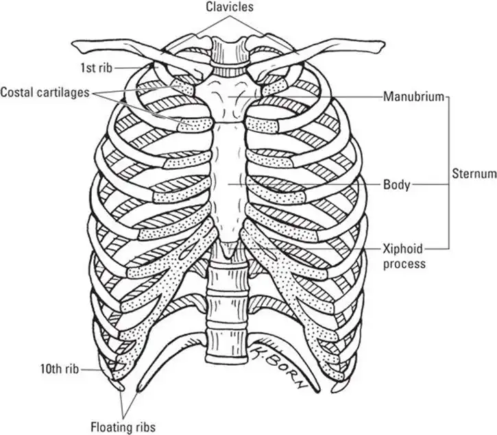
![]() The rib cage (also called the thoracic cage) consists of the thoracic vertebrae, the ribs, and the sternum (see Figure 5-5).
The rib cage (also called the thoracic cage) consists of the thoracic vertebrae, the ribs, and the sternum (see Figure 5-5).
The rib cage is essential for protecting your heart and lungs and for providing a place for the pectoral girdle (scapulae and clavicles) to attach.
You have 12 pairs of bars in your cage. Some of your ribs are true (7), some are false (3), and some are floating (2). All ribs are connected to the bones in your back (the thoracic vertebrae). In the front, true ribs are connected to the sternum (breastbone) by individual costal cartilages (cost- means "rib"); false ribs are connected to the sternum by fused costal cartilage. The last two pairs of ribs are called floating ribs because they remain unattached in the front. The floating ribs give protection to abdominal organs, such as your kidneys, without hampering the space in your abdomen for the intestines.
The sternum (breastbone) has three parts: the manubrium, the body, and the xiphoid (pronounced zi-foid) process. The notch that you can feel at the top center of your chest, in line with your collarbones (the clavicles), is the top of the manubrium. The middle part of the sternum is the body, and the lower part of the sternum is the xiphoid process.
The appendicular skeleton
The appendicular skeleton is made up of the bones and joints of the appendages (upper and lower limbs) and the two girdles that join the appendages to the axial skeleton. We describe each of these categories in the following sections.
Wearing girdles: everybody has two
![]() The word girdle is a verb than means "to encircle." It has nothing to do with that funny undergarment all polite women wore in the early 20th century.
The word girdle is a verb than means "to encircle." It has nothing to do with that funny undergarment all polite women wore in the early 20th century.
The body contains two girdles: the pectoral girdle, which encircles the vertebral column at the top, and the pelvic girdle, which encircles the vertebral column at the bottom. The girdles serve to attach the appendicular skeleton to the axial skeleton.
The pectoral girdle consists of the two clavicles (collarbones) and the two triangle-shaped scapulae (shoulder blades). The scapulae provide a broad surface to which arm and chest muscles attach. Refer to the "Major Bones of the Skeleton" color plate to see the individual parts of the pectoral girdle.
The clavicles are attached to the sternum’s manubrium. Significantly, this is the only point of attachment of the pectoral girdle and the axial skeleton. Because of this relatively weak attachment, the shoulders have a wide range of motion but are prone to dislocation.
Why weight-bearing exercise is good for you
You probably know that aerobic exercise and lifting weights are good for your heart and your muscles, but did you realize it benefits your bones, too? Exercise — especially weight-bearing exercise (such as exercises that use the hips and legs, like walking, running, bicycling, and weight lifting) stimulates remodeling. More remodeling leads to stronger, denser bones. Thus, exercise staves off osteoporosis, which is the loss of bone density and the weakening of bones. Exercising the muscles also exercises the bones and joints, which maintains flexibility and strength. Nothing beats a firm foundation!
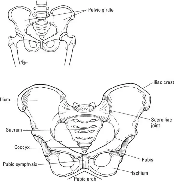
The pelvic girdle (see the pelvis in Figure 5-6) is formed by the hipbones (called coxal bones), the sacrum, and the coccyx (tailbone). The hipbones bear the weight of the body, so they must be strong.
The hipbones (coxal bones) are formed by the ilium, the ischium, and the pubis. The ilia are what you probably think of as your hipbones; they’re the large, flared parts that you can feel on your sides. The part that you can feel at the tip of the ilium is the iliac crest. In your lower back, the ilium connects with the vertebral column at the sacrum; the joint that’s formed is appropriately called the sacroiliac joint — a point of woe for many people with lower back pain.
The ischium is the bottom part of your hip. You have an ischium on each side, within each buttock. You’re most likely sitting on your ischial tuberosity right now. These parts of your hips are also called the sitz bones because they allow you to sit. The ischial tuberosity points outward and is the site where ligaments and tendons from the lower limbs attach. The ischial spine — which is around the area where the ilium and ischium join — is directed inward into the pelvic cavity. The distance between a woman’s ischial spines is key to her success in delivering an infant vaginally (see Chapters 14 and Chapters 15); the opening between the ischial spines must be large enough for a newborn’s head to pass through.
The pubis bones join the right and left hip bones together. They are joined together by a piece of fibrocartilage called the pubic symphysis. Pelvic floor muscles attach to the pelvic girdle at the pubis.
Why women have bigger hips than men
Okay. You know it’s true. Women aren’t built like men. Most men tend to be straight up and down with few curves. Women, on the other hand, are hourglasslike — their hips tend to be wider than men’s hips. In a woman, the iliac bones flare wider than they do in a man. The pubic arch is at an obtuse angle (greater than 90 degrees) where in males it is acute (less than 90 degrees). And the lesser (or true) pelvis of a woman — the ring formed by the pubic bones, ischium, lower part of the ilium, and the sacrum — is wider and more rounded. The lesser pelvis of a man is shaped somewhat like a funnel.
These differences in anatomy have a physiological purpose: Women have babies, and when those babies are ready to be born, they need to pass through a woman’s true pelvis without getting stuck. Other differences also relate to giving birth: The sacrum in women is wider and tilted back more so than in men; the coccyx of women moves easier than it does in men. These two features allow a little more "give" when a baby is passing through the pelvis. When a woman is pregnant, she creates a hormone called relaxin that allows the ligaments connecting the pelvic bones to relax a little. Thus, the bones can spread farther apart and flex a bit during delivery.
All these female characteristics are wonderful for giving birth, but the bones don’t always return to their original positions as readily. The hips tend to stay a bit wider after giving birth. Maybe the bones staying loose and spreading out is a physiological "reward", making delivery of another infant even easier. Too bad female bodies don’t know when that last infant has been delivered so the hips can go back to their pre-pregnant sizes!
Going out on a limb:arms and legs
![]() Your arms and legs are limbs or appendages. The word append means to attach something to a larger body. Your appendages are attached to the axial skeleton by the girdles (see the preceding section).
Your arms and legs are limbs or appendages. The word append means to attach something to a larger body. Your appendages are attached to the axial skeleton by the girdles (see the preceding section).
Giving you a hand with hands (and arms and elbows)
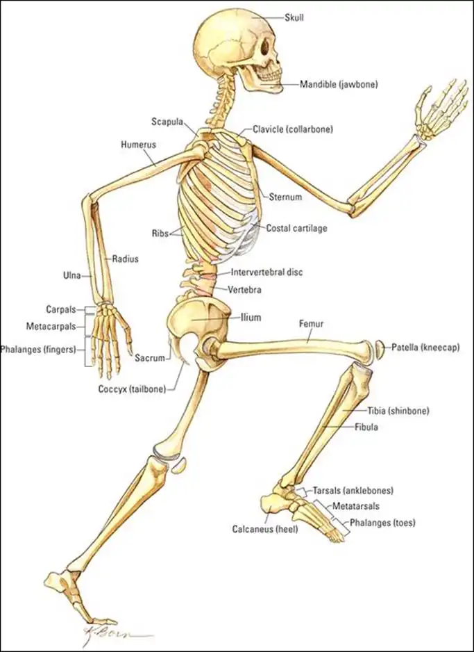
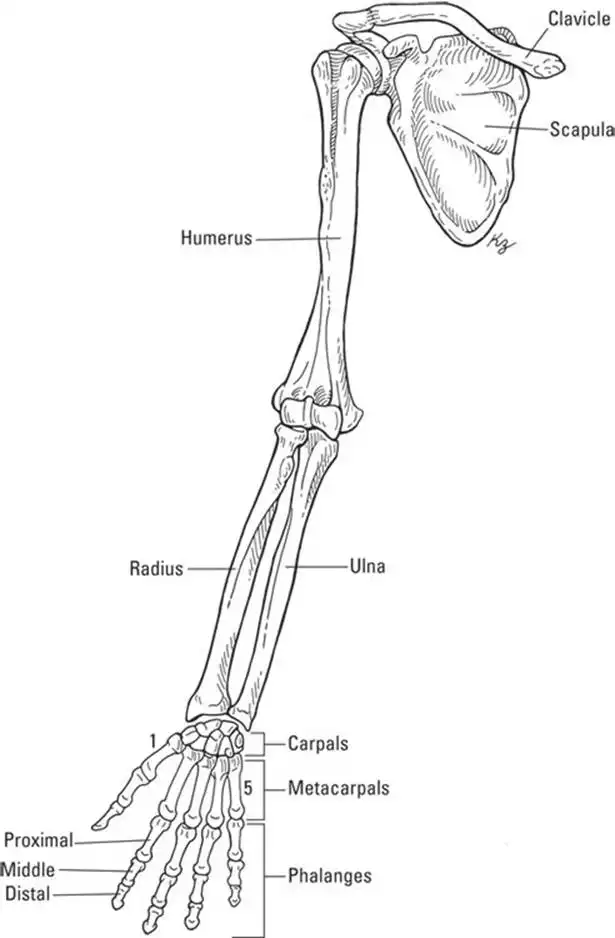
Your upper limb or arm is connected to your pectoral girdle. The bones of your upper limb include the humerus (arm), the radius and ulna (forearm), the carpals of the wrist, and the hand, which is made up of the metacarpal bones and the phalanges (refer to Figure 5-7).
The head (ball at the top) of the humerus connects to the scapula at the glenoid cavity. Muscles that move the arm and shoulder attach to the greater and lesser tubercles, two points near the head. Between the greater and lesser tubercles is the intertubercular groove, which holds the tendon of the biceps muscle to the humerus bone. The humerus also attaches to the deltoid muscle of the shoulder at a point about halfway down called the deltoid tuberosity. The muscle attached to the deltoid tuberosity allows you to raise and lower your arm.
The bones of the forearm attach at the elbow end of your humerus in four different spots:
- Capitulum: Two knobs that allow the radius to articulate (join) with the humerus.
- Trochlea: A pulley-like feature on the humerus that lies next to the capitulum and allows the trochlear notch of the ulna to articulate with the humerus.
- Coronoid fossa: Depression in the humerus that accepts a projection of the ulna bone (called the coronoid process) when the elbow is bent.
- Olecranon fossa: Depression in the humerus that accepts a projection of the ulna (called the olecranon process) when the arm is extended. Fitting, isn’t it?
![]() The radius is the bone on the thumb side of your forearm. When you turn your forearm so that your palm is facing backward, the radius crosses over the ulna so that the radius can stay on the thumb side of your arm. The radius is shorter but thicker than the ulna. The head of the radius is flat like the head of a nail. The ulna is long and thin, and its head is at the opposite end of the bone compared with the head of the radius.
The radius is the bone on the thumb side of your forearm. When you turn your forearm so that your palm is facing backward, the radius crosses over the ulna so that the radius can stay on the thumb side of your arm. The radius is shorter but thicker than the ulna. The head of the radius is flat like the head of a nail. The ulna is long and thin, and its head is at the opposite end of the bone compared with the head of the radius.
Both the radius and ulna connect with the bones of the wrist. The wrist contains eight short bones called the carpal bones. The ligaments binding the carpal bones are very tight, but the numerous bones allow the wrist to flex easily. The eight carpal bones are arranged in two rows. The proximal row (furthest from your fingertips) contain the scaphoid, lunate, triquetrum, and pisiform (from thumb to pinky — note that pisiform and pinky both start with p). The distal row (also from thumb to pinky) contain the trapezium, trapezoid, capitate, and hamate.
The palm of your hand contains five bones called the metacarpals. When you make a fist, you can see the ends of the metacarpals as your knuckles. Your fingers are made up of bones called the phalanges; each finger has three phalanges (phalanx is singular): the proximal phalanx, which joins your knuckle, the middle phalanx, and the distal phalanx, which is the bone in your fingertip. The thumb, though, only has two phalanges, so some people like to argue that it’s not considered a true finger. So you may have eight fingers and two thumbs or ten fingers depending on how you look at it. Regardless, on each hand the thumb is referred to as the first digit.
Getting a leg up on your lower limbs
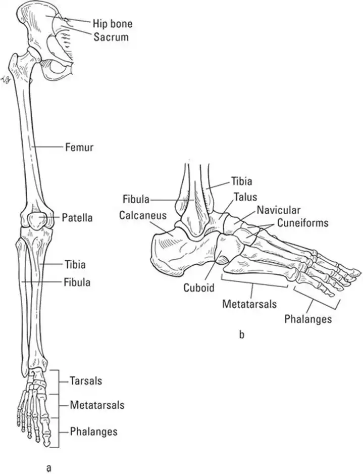
Your lower limb consists of the femur (thigh bone), the tibia and fibula of the leg, the bones of the ankle (tarsals), and the bones of the foot (metatarsals and phalanges; refer to Figure 5-8).
![]() The term phalanges refers to the finger bones and the toe bones.
The term phalanges refers to the finger bones and the toe bones.
The femur is the strongest bone in the body; it’s also the longest. The head of the femur fits into a hollowed out area of the hip bone called the acetabulum. In women, the acetabula (plural) are smaller but spread farther apart than in men. This anatomic feature allows women to have a greater range of movement of the thighs than men. The greater and lesser trochanters of the femur are surfaces to which the muscles of the legs and buttocks attach. Trochanters are large processes found only on the femur. The linea aspera is a ridge along the back of the femur to which several muscles attach.
The femur forms the knee along with bones of the lower leg. The patella (commonly known as the kneecap) articulates with the bottom of the femur. The femur also has knobs (lateral and medial condyles) that articulate with the top of the tibia. The ligaments of the patella attach to the tibial tuberosity. The bottom, inner end of the tibia has a bulge called the medial malleolus, which forms part of the inner ankle.
The tibia, also called the shinbone, is much thicker than the fibula and lies on the medial (inside) portion of the leg. Although the fibula is thinner, it’s about the same length as the tibia. The bottom, outside end of the fibula is the lateral malleolus, which is the bulge on the outside of your ankle.
Your foot is designed in much the same way as your hand. The ankle, or tarsus, which is akin to the wrist, consists of seven tarsal bones. However, only one of those seven bones is part of a joint with a great range of motion — the talus. The talus bone joins to the tibia and fibula and allows for the movements of your ankle. Beneath the talus is the largest tarsal bone, the calcaneus, which is the heel bone. The calcaneus and talus help to support your body weight. The remaining tarsals are the cuboid on the outside, the navicular, and the lateral, intermediate, and medial cuneiforms.
The instep of your foot is akin to the palm of your hand, and just as the hand has metacarpals and phalanges, the foot has metatarsals and phalanges. The ends of the metatarsals on the bottom of your foot form the ball of your foot. As such, the metatarsals also help to support your body weight. Together, the tarsals and metatarsals held together by ligaments and tendons form the arches of your feet. Your toes are also called phalanges, just like your fingers. And, just as your thumbs have only two phalanges, your big toes have only two phalanges. But, the rest of your toes have three: proximal, middle, and distal.
Strong feet, firm foundation
The arches of your feet help to absorb the shock created by the pounding of your feet when walking or running, and they help to distribute your weight evenly to the bones that bear the brunt of it: the calcaneus (heel) and talus (ankle) in each foot. The heel, ankle, and metatarsals bear a significant amount of your body weight and are thus called weight-bearing bones. If the ligaments and tendons that form those arches along with the tarsals and metatarsals weaken, the arches can collapse, resulting in flat feet. A person with flat feet is more prone to damage to the feet bones (such as the metatarsals) because of increased pressure placed on those bones. Examples of damage include bunions, which is a painful displacement of the first metatarsal (the big toe), and heel spurs, which are bony outgrowths on the calcaneus that cause pain when walking. Flat feet also can contribute to knee pain, hip pain, and lower back pain.
See also
- Locating Physiology on the Web of Knowledge
- Chapter 1. Anatomy and Physiology: The Big Picture
- Chapter 2. What Your Body Does All Day
- Chapter 3. A Bit about Cell Biology
- Sizing Up the Structural Layers
- Chapter 4. Getting the Skinny on Skin, Hair, and Nails
- Chapter 5. Scrutinizing the Skeletal System
- Chapter 6. Muscles: Setting You in Motion
- Talking to Yourself
- Chapter 7. The Nervous System: Your Body’s Circuit Board
- Chapter 8. The Endocrine System: Releasing Chemical Messages
- Exploring the Inner Workings of the Body
- Chapter 9. The Cardiovascular System: Getting Your Blood Pumping
- Chapter 10. The Respiratory System: Breathing Life into Your Body
- Chapter 11. The Digestive System: Beginning the Breakdown
- Chapter 12. The Urinary System: Cleaning Up the Act
- Chapter 13. The Lymphatic System: Living in a Microbe Jungle
- Life’s Rich Pageant: Reproduction and Development
- Chapter 14. The Reproductive System
- Chapter 15. Change and Development over the Life Span
- The Part of Tens
- Chapter 16. Ten (Or So) Chemistry Concepts Related to Anatomy and Physiology
- Chapter 17. Ten Phabulous Physiology Phacts
- Supplemental Images

