Expressing your genome
Genes and genetic material
Your anatomical structures are specified in detail, and all your physiological processes are controlled by your very own unique set of genes. Unless you’re an identical twin, this particular set of genes, called your genome, is yours alone, created at the moment your mother’s ovum and your father’s sperm fused. The genome itself (all your genes) is incorporated in the DNA in the nucleus of each and every one of your cells.
![]() The genome consists of many thousands of genes. Early research into the number of individual genes in the human genome has produced varying estimates, from about 23,000 to around 75,000, with estimates toward the lower end of the range predominating. Within any given cell, only a very few of these genes is ever expressed — that is, activated and used in protein formation.
The genome consists of many thousands of genes. Early research into the number of individual genes in the human genome has produced varying estimates, from about 23,000 to around 75,000, with estimates toward the lower end of the range predominating. Within any given cell, only a very few of these genes is ever expressed — that is, activated and used in protein formation.
Traiting you right
Your genes are responsible for your traits. If your genes specify that you’ll grow to 6 feet tall (a trait), they cause bone and tissue to grow until your body reaches that height, and they maintain it thereafter (assuming a favorable environment), until the cells the genes work through age and die. If you have the genes for brown eyes (a trait), your genes direct the production of pigments that color the eyes. And if your genes include those for abundant production of low-density cholesterol, you have a tendency toward atherosclerosis, another trait. In an environment where the available nutritional resources include abundant red meat, this trait could give you trouble. Otherwise, this trait is harmless.
![]() Calling a trait "harmless" may be technically incorrect. Assuming that any trait has some survival-enhancing purpose, at least under some environmental conditions, is probably more correct, even if you don’t know what the purpose is.
Calling a trait "harmless" may be technically incorrect. Assuming that any trait has some survival-enhancing purpose, at least under some environmental conditions, is probably more correct, even if you don’t know what the purpose is.
Gene structure
The physical structure of the DNA molecule, called the DNA double helix, is key to the functioning of genes (refer to Figure 3-4). The DNA double helix can be compared with a ladder or a zipper. In function, it’s more like a system of code.
Code "you"
The nucleotides are the symbols in the genetic code. In modern English, a written word is a code made of certain subunits (letters) set down in a specific order. For example, this, than, then, and ten are different words with different meanings and functions in expressing a thought. A gene is made of certain nucleotides in a certain order. A gene may be a few or many nucleotides in length (imagine a single word 20 pages long), but a given gene is always exactly the same nucleotides in exactly the same order along one strand of DNA. The order of the nucleotides is absolutely crucial: "ACTTAGGCT" is not the same as "ACTAAGGCT." According to the prevailing theory, each gene specifies the construction of one protein molecule. This is called the one gene, one protein model. If the nucleotides are out of order, the protein molecule they make will probably be useless, and the organism’s functioning will likely be impaired, a little or a lot.
![]() Every model used to explain cell biology, including the one gene, one protein model, is subject to change as more information becomes available. But the changes are likely to be minor for understanding basic anatomy and physiology.
Every model used to explain cell biology, including the one gene, one protein model, is subject to change as more information becomes available. But the changes are likely to be minor for understanding basic anatomy and physiology.
Pairing at the molecular level
Remember nucleotide complementary pairs? (If not, see the section "Nucleic acids and nucleotides" earlier in the chapter.) So if a nucleotide is in place on one strand of DNA, and each type of nucleotide binds with only its complementary partner (A with T, C with G, and so on), what do you think is on the other DNA strand? Right! The other nucleotide of the complementary pair. Wherever there’s a G on one strand, there’s a C on the other, and the pair is attached in the middle. And if the two strands become separated (which they do), what do you think will happen? The nucleotides on each strand will attract and hold other molecules of their complementary partners, thus creating two new double-strands identical to the original double-strand. This is how DNA replication occurs.
Synthesizing protein
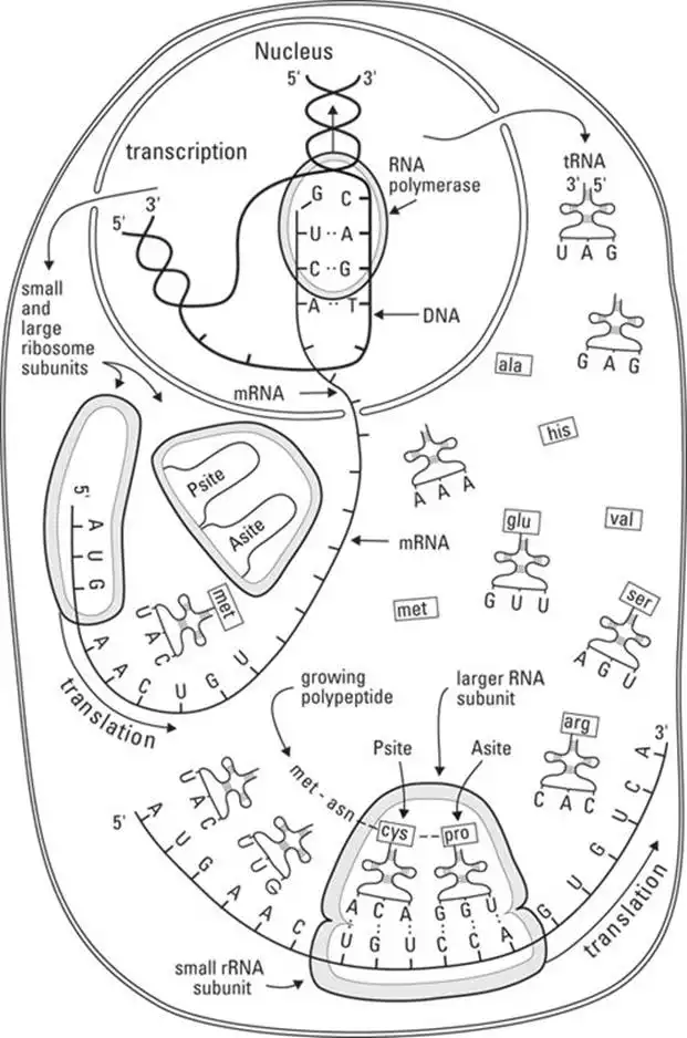
A gene that’s active sends messages to its own cell or to other cells, ordering them to produce molecules of its particular protein, the only one it’s capable of making. The message is sent from DNA through the intermediary of mRNA (messenger RNA), which places an order at the protein factory of the cell and stays around for a while to supervise production. The first part of the process, where DNA "writes the order" in the form of a sequence of nucleotides on mRNA, takes place in the nucleus and is called transcription ("writing across"). The next part of the process, where RNA places the order at the factory, is called translation ("carrying across"). The last part, where the amino acid monomers are sorted out and assembled into the polypeptide, starts in the ribosome. Figure 3-5 shows this process. The finishing touches are put on the protein molecule in the endoplasmic reticulum and the Golgi body. The process from transcription to the last finishing touch on the protein molecule is called gene expression.
![]() Keep in mind the relationship between the genome and gene expression. Your entire genome is contained in identical DNA molecules in the nucleus of every one of your cells; it remains unchanged through your lifetime. Any given gene may be expressed in only a few cells, or only occasionally in a few cells, or only under certain physiological conditions, or possibly never. The totality of gene expression changes every second throughout your lifetime, as quickly as nerves transmit impulses and cells react.
Keep in mind the relationship between the genome and gene expression. Your entire genome is contained in identical DNA molecules in the nucleus of every one of your cells; it remains unchanged through your lifetime. Any given gene may be expressed in only a few cells, or only occasionally in a few cells, or only under certain physiological conditions, or possibly never. The totality of gene expression changes every second throughout your lifetime, as quickly as nerves transmit impulses and cells react.
![]() The terms gene expression, protein synthesis, and transcription-and-translation are essentially the same in meaning, but they’re used in different contexts in cell biology and physiology.
The terms gene expression, protein synthesis, and transcription-and-translation are essentially the same in meaning, but they’re used in different contexts in cell biology and physiology.
The cell cycle
The life cycle of an individual cell is called the cell cycle. The moment of cell cleavage, when a cell membrane grows across the "equator" of a dividing cell, is considered to be the end of the cycle for the mother cell and the beginning of the cycle for each of the daughter cells.
Typically, but by no means universally, interphase is the longest period of the cell cycle. Interphase comes to an end when the cell divides in the process of mitosis. We discuss both these periods of the cell cycle in the following sections.
Uncontrolled cell growth
Our cells have genes that control the cell cycle — that is, how often cells undergo mitosis. Mutations in these genes allow cells to divide unabated. This uncontrolled cell growth is the very definition of cancer. As a result, any tissue in your body has the potential to become cancerous. That most common cancers (worldwide) are lung, breast, colorectal, and prostate.
Cancers are grouped by their origin and the cell type affected. The cells dividing out of control may invade the surrounding tissue, creating a tumor. They may also be knocked loose and take up residence elsewhere, called metastasis. This complicates treatment as you may find a tumor in the prostate gland, but it’s actually cancerous bladder tissue that metastasized there.
Because the cells of each tissue are unique, there is no single cure-all for cancer. Surgery to remove the tumor is often performed if it’s accessible. Radiation treatment may be used to kill the cancers cells in a tumor but kills nearby healthy cells, too. Chemotherapy uses chemicals that target the specific cell type affected. Treatment often involves a combination of all three.
Researchers the world over are trying to find successful, specific treatments that leave the healthy cells unharmed. Unfortunately, we have to battle cancer one tissue type at a time.
Cells that divide, cells that don’t
All cells arise from the division of another cell, but not all cells go on to divide again:
- Zygote: This is the diploid cell that comes into existence when the sex cells (ovum and sperm, both haploid) fuse at conception. Almost immediately, the zygote divides into two somatic cells.
- Somatic cells: These include all the cells of the body except the sex cells — in other words, all the diploid cells of the body. Somatic cells may be relatively differentiated (somewhat specialized), terminally differentiated (they never divide again), or stem cells.
- Stem cells: These are special kinds of rather "generic" somatic cells that divide to produce one new stem cell and one new somatic cell that goes on to differentiate into a particular type of cell in a particular type of tissue. The embryo (organism in the very early stages of development) has very special stem cells, called pluripotent ("many powers") stem cells, which have the ability to give rise to just about any kind of cell an organism needs, given the right chemical environment. When an organism has developed beyond the embryo stage, embryonic stem cells disappear, and other types of stem cells, called multipotent or adult stem cells, arise in particular tissue types and specialize in producing new cells for that tissue. (Turn to Chapter 9 for a description of how stem cells in the bone marrow give rise to many types of blood cells.)
- Sex cells (gametes): These form when specialized somatic cells in the reproductive system divide by a process called meiosis. Meiosis is the only cellular process in the human life cycle that produces haploid cells. See Chapter 14 for more details on sex cells and the processes of meiosis.
Table 3-2 summarizes how different types of cells behave when it comes time to divide.
TABLE 3-2 Dividing Behavior of Different Cell Types
| Organelle | Arise From | Divide? | Give Rise To |
| Zygote | Fusion of two sex cells | Yes | Two somatic cells |
| Somatic cell | Somatic cell or stem cell | Yes or No* | Somatic cells; sex cells** |
| Stem cell | Stem cell | Yes | One specialized somatic cell and one stem cell |
| Sex cell | Somatic cell | No | NA |
* Some somatic cells go on to terminal differentiation and never divide again.
** Sex cells arise from meiosis of certain somatic cells. They are haploid cells and never divide again.
Interphase
Interphase begins when the cell membrane fully encloses the new cell and lasts until the beginning of mitosis or meiosis. The duration of interphase may be anywhere from minutes to decades. Generally speaking (there are always exceptions in cell biology), cells do most of their differentiating and most of their routine metabolizing during interphase. Stem cells grow in size and duplicate organelles during interphase, in preparation for mitosis (more on mitosis in a minute). Some other cells enter mitosis after an extended period of steady-state metabolism. Sometimes, a cell remains in interphase, carrying out its physiological function for years and years until it dies.
DNA replication
DNA replication is an early event in cell division, occurring during interphase, just prior to the beginning of mitosis or meiosis but within the protected space in the nuclear envelope. Maintaining the integrity of the DNA code is absolutely vital.
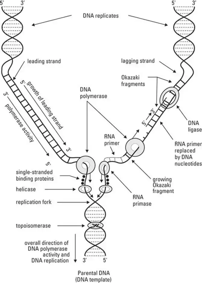
During DNA replication, the double helix must untwist and "unzip" so that the two strands of DNA are split apart. As shown in Figure 3-6, each strand becomes a template for building the new complementary strand. This process occurs a little at a time along a strand of DNA. The entire DNA strand doesn’t unravel and split apart all at once. When the top part of the helix is open, the original DNA strand looks like a Y. This partly open/partly closed area where replication is happening is the replication fork.
![]() In Figure 3-6, the symbols 5' and 3' (read five prime and three prime) indicate the direction in which DNA replication is occurring. The template strand is read in the 3'-to-5' direction. The bases that are complementary to the template strand are added in the 5'-to-3' direction.
In Figure 3-6, the symbols 5' and 3' (read five prime and three prime) indicate the direction in which DNA replication is occurring. The template strand is read in the 3'-to-5' direction. The bases that are complementary to the template strand are added in the 5'-to-3' direction.
Mitosis
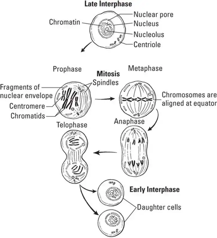
A cell enters a process of mitosis (division) in response to signals from the nucleus. As shown in Figure 3-7, mitosis is a multistage process, proceeding in the following stages:
- Prophase: The nuclear envelope is dismantled, and the duplicated DNA, in the form of chromatin, thickens and coils into chromosomes. Each duplicated chromosome is composed of two identical strands of DNA referred to as chromatids. The chromatids are held together by a protein mass called the centromere. (Note: When the chromatids separate, each is considered a new chromosome.) Cellular structures called centrioles and spindle fibers form and move to the poles of the cell.
- Metaphase: The chromosomes line up to form a perfect row at the center of the cell. The spindle fibers, attached to the centrioles at one end, attach to the chromosomes. At this point, 92 chromatids are in a double set of 46 chromosomes.
- Anaphase: The centromere is split by enzymatic activity, and the chromatids are pulled apart by the spindle fibers toward one of the centrioles: 46 to one, 46 to the other. Following this movement, the chromosomes are referred to as daughter chromosomes, and the set at one pole is identical to the set at the opposite pole. But the cell isn’t quite ready to divide yet.
- Telophase: A fresh nuclear membrane is reassembled around each set of chromosomes. The spindles dissolve, which frees the daughter chromosomes.
At this point, when each of the two identical nuclei is at one pole of the cell, mitosis is technically over. However, the cell’s cytoplasm still has to actually split apart into two masses, a process called cytokinesis. The center of the mother cell indents and squeezes the cell membrane across the cytoplasm until two separate cells are formed. The two daughter cells are then in interphase and go on to differentiation or not, depending on the instructions to the cell from the genome.
See also
- Locating Physiology on the Web of Knowledge
- Chapter 1. Anatomy and Physiology: The Big Picture
- Chapter 2. What Your Body Does All Day
- Chapter 3. A Bit about Cell Biology
- Sizing Up the Structural Layers
- Chapter 4. Getting the Skinny on Skin, Hair, and Nails
- Chapter 5. Scrutinizing the Skeletal System
- Chapter 6. Muscles: Setting You in Motion
- Talking to Yourself
- Chapter 7. The Nervous System: Your Body’s Circuit Board
- Chapter 8. The Endocrine System: Releasing Chemical Messages
- Exploring the Inner Workings of the Body
- Chapter 9. The Cardiovascular System: Getting Your Blood Pumping
- Chapter 10. The Respiratory System: Breathing Life into Your Body
- Chapter 11. The Digestive System: Beginning the Breakdown
- Chapter 12. The Urinary System: Cleaning Up the Act
- Chapter 13. The Lymphatic System: Living in a Microbe Jungle
- Life’s Rich Pageant: Reproduction and Development
- Chapter 14. The Reproductive System
- Chapter 15. Change and Development over the Life Span
- The Part of Tens
- Chapter 16. Ten (Or So) Chemistry Concepts Related to Anatomy and Physiology
- Chapter 17. Ten Phabulous Physiology Phacts
- Supplemental Images


