Differentiating the three types of muscle tissue
Talking about tissue types
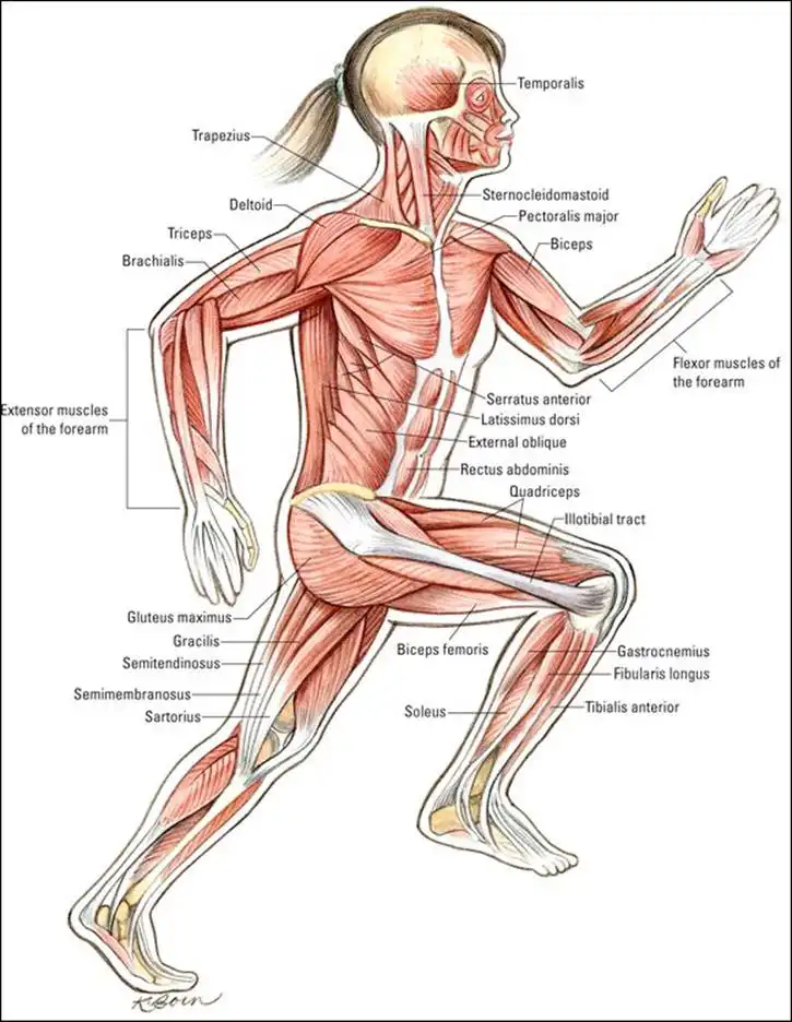
A "muscle tissue type" is not the same as "a muscle". Your left bicep is a muscle; in all, you have hundreds of named muscles. (Refer to the "Naming the Skeletal Muscles" section later in the chapter and to the "Muscular System" color plate in the center of the book.) There are only three muscle tissue types: skeletal muscle tissue, cardiac muscle tissue, and smooth muscle tissue.
Defining unique features of muscle cells
Your muscle tissue is made up of cells that are different from the other cells of your body. These cells are so unique that they’re even different from each other, based on the type of muscle tissue they belong to. The three muscle types are distinguishable anatomically by their characteristic cells and structures and physiologically as voluntary or involuntary.
Muscle cells feature these characteristics:
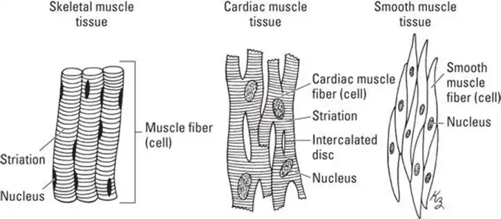
- Single or multiple nuclei: Cardiac muscle cells and smooth muscle cells have one nucleus apiece, like most other cells. Skeletal muscle cells (referred to as fibers) are multinucleate, meaning numerous nuclei are found within one cell membrane. Skeletal muscle cells don’t grow extra nuclei; during the development of skeletal muscle tissue, numerous skeletal muscle cells merge into one large cell, and most of the nuclei are retained within one continuous cell membrane, along with most of the mitochondria.
- Striation: Skeletal muscle is striated, meaning that, under a microscope, alternating light and dark bands are visible in the fiber (muscle cell). Striation is the result of the structures inside the skeletal muscle cells (see the "Skeletal Muscle" section later in the chapter) that carry out the mechanism of contraction called the sliding filament model. (See the "Getting a Grip on the Sliding Filament" section later in the chapter). Cardiac muscle cells are striated as well, and they also contract by a variation of the sliding filament model. Smooth muscle cells are not striated in appearance but do follow a version of the sliding filament model.
See Figure 6-1 to get an idea of how muscle cells and tissues are similar and different.
Muscle cells can also be categorized by the type of contraction they perform. Smooth and cardiac muscle cells are involuntary, meaning their contraction is initiated and controlled by parts of the nervous system that are far from the conscious level of the brain (the autonomic nervous system). You have no practical way to consciously control, or even become aware of, the smooth muscle contractions in your stomach that are grinding up this morning’s muffin. The involuntary contractions that cause your heartbeat aren’t even under the nervous system’s control, as we discuss in Chapter 9.
Skeletal muscle is classified as voluntary because you make a decision at the conscious level to move the muscle. At least you do sometimes — you decide to reach for a doorknob and turn it, for example, and your muscles carry out the command from your brain to do so.
But note this: If the doorknob is charged with static electricity, your arm pulls your hand away before you’re even consciously aware of being zapped. This somatic reflex arc is still classified as voluntary movement, however, because it involves skeletal muscle, controlled by the somatic (voluntary) nervous system.
![]() Not everything in anatomy and physiology makes sense at first. Just remember that skeletal muscle is classified as voluntary.
Not everything in anatomy and physiology makes sense at first. Just remember that skeletal muscle is classified as voluntary.
Table 6-1 sums up the characteristics and classifications of muscle cells.
TABLE 6-1 Cell Characteristics of Muscle Cells
| Muscle | Skeletal | Cardiac | Smooth |
| Multinucleate | Yes | No | No |
| Striated | Yes | Yes | No |
| Voluntary | Yes | No | No |
Skeletal muscle
Skeletal muscle tissue is, essentially, bundles of fibers bundled together. Like fibrous material of every kind, skeletal muscle tissue gets its strength from assembling individual fibers together into strands, and then bundling and rebundling the strands. Two properties make this particular fibrous material very special: The strands are made of protein, and they renew and repair themselves constantly.
At the cellular level
Individual muscle cells, which physiologists call fibers, are slender cylinders that sometimes run the entire length of a muscle. Each fiber (cell) has many nuclei located along its length and close to the cell membrane, which is called the sarcolemma in skeletal muscle fibers. Outside the sarcolemma is a lining called the endomysium, a type of connective tissue, which houses capillaries and nerves.
Muscle spindles are specialized skeletal muscle fibers that are wrapped with nerve fibers. Figure 6-2 shows how skeletal muscle is connected to the nervous system. Spindles are distributed throughout the muscle tissue and provide sensory information to the central nervous system. Motor neurons transmit impulses to trigger a muscle fiber to contract. Each fiber must be stimulated individually by a neuron at its motor end plate. However, a single motor neuron can stimulate numerous fibers, forming a motor unit. Large motor units (one neuron, numerous fibers) allow for gross motor skills like walking and lifting. Small motor units (one neuron, few fibers) provide fine motor skills like grasping and handwriting.
Bundled within the muscle fibers are myofibrils (refer to Figure 6-2). The myofibrils are composed of sarcomeres, which are distinct units arranged linearly (end to end) along the length of the myofibril. A sarcomere is the functional unit of muscle contraction. (Refer to the "Getting a Grip on the Sliding Filament Model" section later in the chapter for more on muscle contraction within sarcomeres.)
At the tissue level
Muscle fibers are bound together into bundles called fascicles. Each fascicle is bound by a connective-tissue lining called a perimysium. Spindle fibers are distributed throughout each fascicle. The fascicles are then bound together to form a muscle, a discrete assembly of skeletal muscle tissue, like the biceps brachii (your biceps), with a connective-tissue wrapper called an epimysium holding the whole package together.
Tendons — ropy extensions of the connective tissue covering the skeletal bones — weave into the epimysium, holding the muscle firmly to the bone (see Chapter 5 for more on connectivity in the skeletal system). Muscles connect to other muscles using aponeuroses, a connective tissue similar to a tendon but broad and flat.
![]() How many ways can you say "fiber"? Anatomists need them all when they’re talking about the muscular system. Make sure you’re thinking at the right level of organization (subcellular, cellular, or tissue) when you see these terms: filament, myofibril, fiber, and fascicle.
How many ways can you say "fiber"? Anatomists need them all when they’re talking about the muscular system. Make sure you’re thinking at the right level of organization (subcellular, cellular, or tissue) when you see these terms: filament, myofibril, fiber, and fascicle.
Working together: synergists and antagonists
Groups of skeletal muscles that contract simultaneously to move a body part are said to be synergistic. The muscle that does most of the moving is the prime mover. The muscles that help the prime mover achieve a certain body movement are synergists. When you move your elbow joint, the bicep is the prime mover and the brachioradialis stabilizes the joint, thus aiding the motion.
Antagonistic muscles also act together to move a body part, but one group contracts while the other releases, a kind of push-pull. One example is flexing your arm. When you bend your forearm up toward your shoulder, your biceps muscle contracts, performing a concentric contraction. In the meantime, the triceps muscle in the back of your arm relaxes, performing an eccentric contraction. The actions of the biceps and triceps muscles are opposite, but you need both actions to allow you to flex your arm, which is why they are both, confusingly, referred to as contractions. Antagonistic actions lower your arm, too: the biceps relaxes, and the triceps contracts.
Cardiac muscle
The heart has its own very special type of muscle tissue, called cardiac muscle. The cells (fibers) in cardiac muscle contain one nucleus (they’re uninucleated) and are cylindrical; they may be branched in shape. Unlike skeletal muscle, where the fibers lie alongside one another, cardiac muscle fibers interlock, which promotes the rapid transmission of the contraction impulse throughout the heart. Cardiac muscle cells are striated, like skeletal muscle cells, and cardiac muscle contraction is involuntary, like smooth muscle contraction. Cardiac muscle fibers contract in a way very similar to skeletal muscle fibers, by a sliding filament mechanism (more on that in a minute).
Cardiac muscle tissue is on the job, day and night, from before birth to the moment of death. The cardiac muscle cells contract regularly and simultaneously hundreds of millions of times throughout your lifetime. When cardiac muscle tissue gives up, the game is over.
Unlike skeletal muscle and smooth muscle, contraction of the heart muscle is autonomous, which means it occurs without stimulation by a nerve. In between contractions, the fibers relax completely (see Chapter 9).
Smooth muscle
Smooth muscle tissue is found in the walls of organs and structures of many organ systems, including the digestive system, the urinary system, the respiratory system, the cardiovascular system, and the reproductive system. Smooth muscle tissue is fundamentally different from skeletal muscle tissue and cardiac muscle tissue in terms of cell structure and physiological function. However, smooth muscle sarcomeres are similar.
Smooth muscle fibers (cells) are fusiform (thick in the middle and tapered at the ends) and arranged to form sheets of tissue. Smooth muscle cells aren’t striated. However, smooth muscle contractions utilize the same sliding filament mechanism as skeletal muscle cells (see the next section for more).
Smooth muscle contraction is typically slow, strong, and enduring. Smooth muscle can hold a contraction longer than skeletal muscle. In fact, some smooth muscles, notably the sphincters, are in a constant state of contraction. Childbirth is among the few occasions in life when humans (some humans, anyway) consciously experience smooth muscle contraction (although they don’t consciously control it).
See also
- Locating Physiology on the Web of Knowledge
- Chapter 1. Anatomy and Physiology: The Big Picture
- Chapter 2. What Your Body Does All Day
- Chapter 3. A Bit about Cell Biology
- Sizing Up the Structural Layers
- Chapter 4. Getting the Skinny on Skin, Hair, and Nails
- Chapter 5. Scrutinizing the Skeletal System
- Chapter 6. Muscles: Setting You in Motion
- Talking to Yourself
- Chapter 7. The Nervous System: Your Body’s Circuit Board
- Chapter 8. The Endocrine System: Releasing Chemical Messages
- Exploring the Inner Workings of the Body
- Chapter 9. The Cardiovascular System: Getting Your Blood Pumping
- Chapter 10. The Respiratory System: Breathing Life into Your Body
- Chapter 11. The Digestive System: Beginning the Breakdown
- Chapter 12. The Urinary System: Cleaning Up the Act
- Chapter 13. The Lymphatic System: Living in a Microbe Jungle
- Life’s Rich Pageant: Reproduction and Development
- Chapter 14. The Reproductive System
- Chapter 15. Change and Development over the Life Span
- The Part of Tens
- Chapter 16. Ten (Or So) Chemistry Concepts Related to Anatomy and Physiology
- Chapter 17. Ten Phabulous Physiology Phacts
- Supplemental Images


