Looking at anatomy: planes, regions, and cavities
Looking at the body from the proper perspective
Remember that story about a friend of a friend that went in to have a foot amputated only to awaken from surgery to find they removed the wrong one? This story highlights the need for a consistent perspective to go with the jargon. Terms that indicate direction make no sense if you’re looking at the body the wrong way. You likely know your right from your left, but ignoring perspective can get you all mixed up. This section shows you the anatomical position, planes, regions, and cavities, as well as the main membranes that line the body and divide it into major sections.
Getting in position
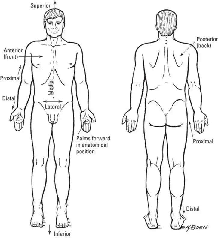
Stop reading for a minute and do the following: Stand up straight. Look forward. Let your arms hang down at your sides and turn your palms so they’re facing forward. You are now in anatomical position (see Figure 1-1). Unless you are told otherwise, any reference to location (diagram or description) assumes this position. Using anatomical position as the standard removes confusion.
The following list of common anatomical descriptive terms (direction words) that appear throughout this and every other anatomy book may come in handy:
- Right: Toward the patient’s right
- Left: Toward the patient’s left
- Anterior/ventral: Front, or toward the front of the body
- Posterior/dorsal: Back, or toward the back of the body
- Medial: Toward the middle of the body
- Lateral: On the side or toward the side of the body
- Proximal: Nearer to the point of attachment or the trunk of the body
- Distal: Farther from the point of attachment or the trunk of the body (think "distance")
- Superficial: Nearer to the surface of the body
- Deep: Farther from the surface of the body
- Superior: Above or higher than another part
- Inferior: Below or lower than another part
![]() Notice that this list of terms is actually a series of pairs. Learning them as pairs is more effective and useful.
Notice that this list of terms is actually a series of pairs. Learning them as pairs is more effective and useful.
Dividing the anatomy
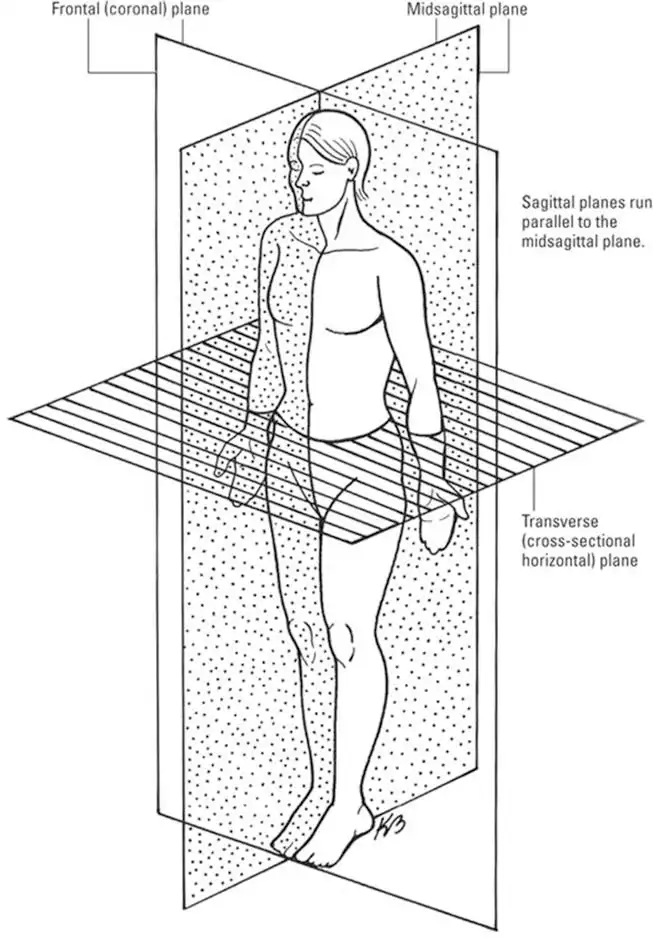
If you’ve taken geometry, you know that a plane is a flat surface and that a straight line can run between two points on that flat surface. Geometric planes can be positioned at any angle. In anatomy, generally three planes are used to separate the body into sections. Figure 1-2 shows you what each plane looks like. The reason for separating the body with imaginary lines — or making actual cuts referred to as sections — is so that you know which half or portion of the body or organ is being discussed. When identifying or comparing structures, you need to know your frame of reference. The anatomical planes are as follows:
- Frontal plane: Divides the body or organ into anterior and posterior portions — think front and back.
- Sagittal plane: Divides the body or organ lengthwise into right and left sections. If the vertical plane runs exactly down the middle of the body, it’s referred to as the midsagittal plane.
- Transverse plane: Divides the body or organ horizontally, into superior and inferior portions — think top and bottom. Diagrams from this perspective can be quite disorienting. You can think of the body like a music box that has a top that opens on a hinge. The transverse plane is where the music box top separates from the bottom of the box. Imagine that you open the box by lifting the lid and are looking down at the contents.
![]() Anatomical planes do not always create two equal portions and can "pass through" the body at any angle. The three planes provide an important reference but don’t expect the structures of the body, and especially the joints, to line up or move along the standard planes and axes.
Anatomical planes do not always create two equal portions and can "pass through" the body at any angle. The three planes provide an important reference but don’t expect the structures of the body, and especially the joints, to line up or move along the standard planes and axes.
Mapping out your regions
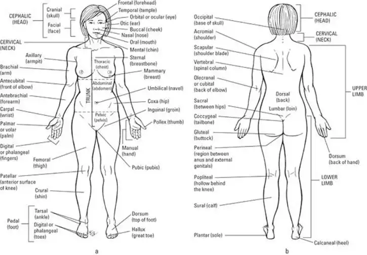
The anatomical planes orient you to the human body, but regions (shown in Figure 1-3) compartmentalize it. Just like on a map, a region refers to a certain area. The body is divided into two major portions: axial and appendicular. The axial body runs right down the center (axis) and consists of everything except the limbs, meaning the head, neck, thorax (chest and back), abdomen, and pelvis. The appendicular body consists of appendages, otherwise known as upper and lower extremities (which you call arms and legs).
Here’s a list of the axial body’s main regions:
- Head and neck
- Cephalic (head)
- Cervical (neck)
- Cranial (skull)
- Frontal (forehead)
- Nasal (nose)
- Occipital (base of skull)
- Oral (mouth)
- Orbital/ocular (eyes)
- Thorax
- Axillary (armpit)
- Costal (ribs)
- Deltoid (shoulder)
- Mammary (breast)
- Pectoral (chest)
- Scapular (shoulder blade)
- Sternal (breastbone)
- Vertebral (backbone)
- Abdomen
- Abdominal (abdomen)
- Gluteal (buttocks)
- Inguinal (bend of hip)
- Lumbar (lower back)
- Pelvic (area between hipbones)
- Perineal (area between anus and external genitalia)
- Pubic (genitals)
- Sacral (end of vertebral column)
Here’s a list of the appendicular body’s main regions:
- Upper extremity
- Antebrachial (forearm)
- Antecubital (inner elbow)
- Brachial (upper arm)
- Carpal (wrist)
- Cubital (elbow)
- Digital (fingers/toes)
- Manual (hand)
- Palmar (palm)
- Lower extremity
- Crural (shin, front of lower leg)
- Femoral (thigh)
- Patellar (front of knee)
- Pedal (foot)
- Plantar (arch of foot)
- Popliteal (back of knee)
- Sural (calf, back of lower leg)
- Tarsal (ankle)
Casing your cavities
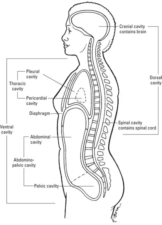
If you remove all the internal organs, the body is empty except for the bones and other tissues that form the space where the organs were. Just as a dental cavity is a hole in a tooth, the body’s cavities are "holes" where organs are held (see Figure 1-4). The two main cavities are the dorsal cavity and the ventral cavity.
The dorsal cavity consists of two cavities that contain the central nervous system. The first is the cranial cavity, the space within the skull that holds your brain. The second is the spinal cavity (or vertebral cavity), the space within the vertebrae where the spinal cord runs through your body.
The ventral cavity is much larger and contains all the organs not contained in the dorsal cavity. The ventral cavity is divided by the diaphragm into smaller cavities: the thoracic cavity, which contains the heart and lungs, and the abdominopelvic cavity, which contains the organs of the abdomen and the pelvis. The thoracic cavity is divided into the right and left pleural cavities (lungs) and the pericardial cavity (heart). The abdominopelvic cavity is also subdivided. The abdominal cavity contains organs such as the stomach, liver, spleen, and most of the intestines. The pelvic cavity contains the reproductive organs, the bladder, the rectum, and the lower portion of the intestines.
Additionally, the abdomen is divided into quadrants and regions. The mid-sagittal plane and a transverse plane intersect at an imaginary axis passing through the body at the umbilicus (navel or belly button). This axis divides the abdomen into quadrants (four sections). Putting an imaginary cross on the abdomen creates the right upper quadrant, left upper quadrant, right lower quadrant, and left lower quadrant. Physicians take note of these areas when a patient describes symptoms of abdominal pain.
The regions of the abdominopelvic cavity include the following:
- Epigastric: The central part of the abdomen, just above the navel
- Hypochondriac: Doesn’t moan about every little ache and illness but lies to the right and left of the epigastric region and just below the cartilage of the rib cage (chondral means "cartilage", and hypo- means "below")
- Umbilical: The area around the umbilicus
- Lumbar: Forms the region of the lower back to the right and left of the umbilical region
- Hypogastric: Below the stomach and in the central part of the abdomen, just below the navel
- Iliac: Lies to the right and left of the hypogastric regions near the hipbones
See also
- Locating Physiology on the Web of Knowledge
- Chapter 1. Anatomy and Physiology: The Big Picture
- Chapter 2. What Your Body Does All Day
- Chapter 3. A Bit about Cell Biology
- Sizing Up the Structural Layers
- Chapter 4. Getting the Skinny on Skin, Hair, and Nails
- Chapter 5. Scrutinizing the Skeletal System
- Chapter 6. Muscles: Setting You in Motion
- Talking to Yourself
- Chapter 7. The Nervous System: Your Body’s Circuit Board
- Chapter 8. The Endocrine System: Releasing Chemical Messages
- Exploring the Inner Workings of the Body
- Chapter 9. The Cardiovascular System: Getting Your Blood Pumping
- Chapter 10. The Respiratory System: Breathing Life into Your Body
- Chapter 11. The Digestive System: Beginning the Breakdown
- Chapter 12. The Urinary System: Cleaning Up the Act
- Chapter 13. The Lymphatic System: Living in a Microbe Jungle
- Life’s Rich Pageant: Reproduction and Development
- Chapter 14. The Reproductive System
- Chapter 15. Change and Development over the Life Span
- The Part of Tens
- Chapter 16. Ten (Or So) Chemistry Concepts Related to Anatomy and Physiology
- Chapter 17. Ten Phabulous Physiology Phacts
- Supplemental Images

