Endoscopic Treatment of the C2 (axis) fractures
Description of the operation method
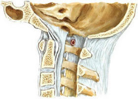
If the fractures of the axial vertebrae are in the lower or middle region, then they are often not stable and require osteosynthesis.
In this case, there have been two operational methods so far:
- Access from the back of the neck (posterior) allows for osteosynthesis between the first and second cervical vertebrae using a bone graft from the iliac crest. This method has paid off and provides high stability. One significant drawback of this method is the complete limitation of movement between the first and second cervical vertebrae, which in turn leads to a loss of rotational ability by 30–40%.
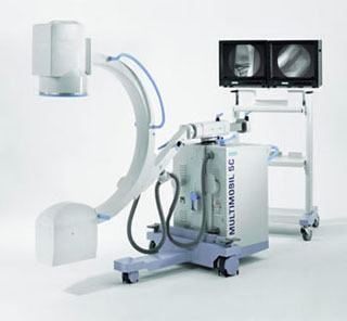
- Osteosynthesis from the front of the neck (anterior). With this method, a single screw is inserted into the axis of the axial vertebra. This ensures a flawless osteosynthesis of the bone without subsequent loss of rotational ability. From the point of view of surgery, this method is less laborious compared to other surgical methods. This, however, requires the simultaneous use of two video amplifiers, the number of which is often limited. Also, the place where the screw is inserted is installed using a video amplifier, and at the same time, the surgeon does not have a direct view of this place, since the original entrance is very remote from the insertion point.
Recommended surgical method for endoscopically assisted treatment of axial vertebral fractures
To limit the use of an X-ray video amplifier, an AP amplifier (posterior/anterior) can be used. In this case, the image is recorded only after positioning the retractor blade. Lateral perspective is used throughout the entire intervention to ensure perfect screw control. The AP image (back and front image) is needed to check the position of the K-string before installing the screw.
We prefer fluorescent navigation for this action. The excellent resolution due to the X-ray conducting retractor blade and the good positioning of the screw achieved by navigation in combination with the lateral X-ray image increases the reliability of this method. We, therefore, recommend video-assisted surgery, which has two advantages:
- on the one hand, the possibility of sufficient illumination of the operating field
- on the other hand, easier identification of the middle of the spine to install the osteosynthesis screw
The retractor blade is made of X-ray conductive metal material to achieve enhanced video amplifier imaging. The end of the retractor blade is curved to provide visibility of the surgical site as well as sufficient subsequent optical control. It also uses a straight endoscope with a 0 ° viewing direction. The retractor blade can be temporarily anchored to the bone using two metal rods. The handle is in a vertical position in relation to the axis, which does not restrict the movement of the surgeon.
See also
- B-Twin sliding implant for spinal fixation
- Endoscopic mcrodiscectomy for herniated or protruded intervertebral discs
- Endoscopic treatment of the C2 (axis) fractures
- Epiduroscopy
- Laser reconstruction of intervertebral discs
- Laser reconstruction of intervertebral discs
- Percutaneous posterolateral foraminoscopy
- Retractor with a lighting system for a herniated disc
- Spinoscope for the treatment of herniated intervertebral disc and stenotic intervertebral foramen
- Technique of sampling and blending sponge layer (spongiosa)
- Transforaminal endoscopic discectomy (TFED)
- Trocars for endoscopic spine surgery
- Vertebroplasty and kyphoplasty with the "SKy" intravertebral bone expander system


