Endoscopic Microdiscectomy for Herniated or Protruded Intervertebral Discs
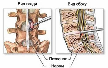
Description of the operation for endoscopic removal of hernia or protrusion of the intervertebral disc of the lumbar spine
Hernia or protrusion of the intervertebral disc of the lumbar spine is very common and relatively rarely requires surgical intervention (operation) to remove it. Nevertheless, more than 200,000 operations are performed annually in the United States, and 20,000 operations are performed in Germany to remove an intervertebral hernia or protrusion of the intervertebral disc of the lumbar spine. The usual surgical treatment for a herniated or protruded intervertebral disc of the lumbar spine is discectomy (removal of protruding disc fragments from the spinal canal, followed by revision of the disc cavity) with a posterior approach under local conduction (epidural) anesthesia or general endotracheal anesthesia.
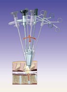
The use of an endoscope during an operation to remove a hernia or protrusion of the intervertebral disc of the lumbar spine allows you to maintain the same access and the same technique for performing a spinal operation, however, the size of the skin incision and the entire access during the operation is significantly reduced.
This method of surgery has the same advantages as microscopic or conventional open (abdominal) discectomy, however, the direct postoperative consequences are not so complicated, which provides a faster and more relaxed rehabilitation of the patient and the resumption of the previous work activity.
In addition, the small size of the skin incision during surgery for a hernia or protrusion of the intervertebral disc of the lumbar spine has certain aesthetic advantages of the spine. The minimal consequences after the patient being operated on to remove a herniated disc are the low trauma of the access and quick rehabilitation after the removal of the fallen out fragments of the disc itself.
Surgical technique of surgery to remove a hernia or protrusion of the intervertebral disc of the lumbar spine
Under X-ray control, using a localization frame, the damaged intervertebral lumbar spine and the direction of approach to it are indicated. A small skin incision over the projection of the damaged disc of the lumbar spine from 10 to 15 mm, depending on the completeness of the operated patient.
The lumbar aponeurosis is dissected with scissors and the paravertebral muscle mass is dissected from the side of the disc herniation. A special operating tube is inserted with an obturator, the visible intralaminar surface is cleaned by means of disc forceps, an endoscopic device with suction is inserted, and further intervention continues under visual control using a video camera.
Resection of parts of the upper arch of the vertebra (laminotomy) is performed so that the upper part of the ligamentum flavum (ligamentum flavum) can be folded back. A part of the articular mass (intervertebral joint) is also removed in order to release the outer edge of the "dural funnel" and the beginning of the affected nerve root.
With the help of a special hook and an elevator, the nerve root is dissected, providing access to a hernia or protrusion of the intervertebral disc. The presence of several instrumental canals when removing a herniated disc of the lumbar spine makes it possible to better manipulate the anatomical elements in the canal. The hook, with the help of which the nerve roots move apart, allows you to open a hernia or protrusion of the intervertebral disc and operate it without the risk of damaging the nerve structures in the lumen of the spinal canal.
Depending on the specific case, after removal of the prolapsed fragments of the disc herniation, a subsequent microdiscectomy can be performed. The disc cavity is washed after the disc herniation and microdiscectomy surgery. Hemostasis (stopping bleeding) is usually achieved by simple plugging or by bipolar coagulation of bleeding epidural veins. When removing the endoscope, the hemostasis of the muscle mass of the spine must be performed very carefully.
Finding the endoscope inside the spinal canal of the lumbar spine provides a very wide angle of vision for the neurosurgeon and allows the detection of wandering fragments of the herniated intervertebral disc (sequesters or parts of hernia that have been laced under the posterior longitudinal ligament).
The operation ends with the imposition of an intradermal suture and a waterproof bandage. After the operation of endoscopic removal of the herniated disc of the lumbar or cervical spine, immediate rehabilitation follows.
See also
- B-Twin sliding implant for spinal fixation
- Endoscopic mcrodiscectomy for herniated or protruded intervertebral discs
- Endoscopic treatment of the C2 (axis) fractures
- Epiduroscopy
- Laser reconstruction of intervertebral discs
- Laser reconstruction of intervertebral discs
- Percutaneous posterolateral foraminoscopy
- Retractor with a lighting system for a herniated disc
- Spinoscope for the treatment of herniated intervertebral disc and stenotic intervertebral foramen
- Technique of sampling and blending sponge layer (spongiosa)
- Transforaminal endoscopic discectomy (TFED)
- Trocars for endoscopic spine surgery
- Vertebroplasty and kyphoplasty with the "SKy" intravertebral bone expander system


