Neuroendoscope
Neuroendoscope
Endoscopy in neurosurgery was developed by W. Dandy and V.I.Mixter at the beginning of the twentieth century and, thanks to the technical improvement carried out in the 60s by G. Guiot, is also used in modern medicine. In 1996, this increasingly popular method was used primarily for the four important indications listed below.
Treatment of obstructive hydrocephalus
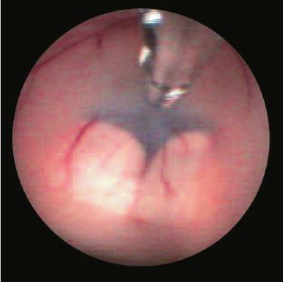
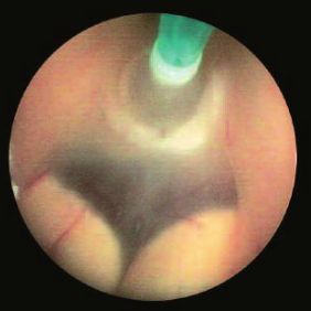
Ventriculocisternostomy covers approximately 80% of neuroendoscopy indications. This intervention provides an effective treatment for hydrocephalus obstruction below the anterior part of the 3rd ventricle (posterior part of the 3rd ventricle, Regio pinealis, Sylvian aqueduct, fourth ventricle, foramen of Magendie). Obstructive hydrocephalus accounts for approximately 20% of all hydrocephalus. A condition for ventriculocisternostomy is the active function of the resorption zones of the cerebrospinal fluid. Endoscopic control ensures that the fundus of the 3rd ventricle is completely safe to open in front of the Corpora mamillaria, thereby creating a direct connection between the dilated spaces and the pericrebral subarachnoid spaces.
Also, neuroendoscopy allows the treatment of occlusive hydrocephalus without the need for multiple bypass systems.
Marsupialization of arachnoid cysts
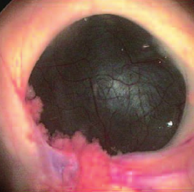
Neuroendoscopy allows the creation of multiple connections between arachnoid cysts and ventricular and/or skull base cavities. Ventriculocystocystomy for suprasellar and temporal arachnoid cysts is the most common indication.
Treatment of colloidal cysts of the 3rd ventricle
Neuroendoscopy makes it possible to puncture colloidal cysts of the anterior part of the 3rd ventricle with optical control in a way that is gentle on the surrounding structures (Plexus choroidei, Venae choroideae, Vena thalamostriata, and Vena septi, as well as the anterior fornix). Microscissors can be used to open the sidewall of the cyst wide to facilitate emptying of the cyst. The remaining fragments of the lateral wall are then treated with electrocautery to avoid relapse.
Biopsy
Neuroendoscopy allows biopsy of tumors with intraventricular development, as well as biopsy of the lateral walls of cystic tumors, while the negligible size of the fragments taken requires multiple samples in each case. In case of bleeding, hemostasis is possible through coagulation.
Fixation and control of the neuroendoscope
- Endoscope fixation: It is not recommended to hold the endoscope in your hands due to possible interfering movements, especially when using the instrument in the operating channel. The use of a hinged arm allows the neuroendoscope to be fixed in any desired position at any time. This leaves your hands completely free for safe working with tools. The locking system, which simultaneously unlocks the hinges of the arm, allows unhindered control of the device for an easy and quick change of the neuroendoscope position.
- Endoscope control: Most applications of endoscopy involve the intervention of the dilated ventricular system using its anterior sheath. This operation, which is identical to a simple ventricular puncture, is carried out without problems with free hands, while the neuroendoscope can be fixed at any time using a hinged arm. For moderate expansion and all other points of impact, stereotactic control of the neuroendoscope is strongly recommended. The articulated arm is designed to be used in conjunction with a stereotactic frame. Once the neuroendoscope has been brought into the desired position and/or the anatomical structures depicted are identified, the stereotactic frame arch can be removed and the intervention can be continued with only the articulated arm.
See also
- Diagnostic optics for endoscopically assisted mcroneurosurgery
- Endonasal endoscopic removal of neoplasms in the nose and paranasal cavities
- Endoscopic pituitary surgery
- Endoscopic transnasal neurosurgery
- Endoscopically assisted microneurosurgery (EAM)
- Ethmoid-pterygo-sphenoidal endoscopic approach (EPSea)
- Extended endonasal approach (EEA) in skull base surgery
- Minimally invasive microscopic surgery of the lateral skull base
- Neuroendoscope:
- Neuroendoscopy
- Neuroendoscopy in pediatrics
- Optical retractor for endoscopically assisted collection a. temporalis superficialis and cranioplasty
- Transphenoidal skull base microsurgery


