Endoscopically Assisted Microneurosurgery (EAM)
Endoscopically Assisted Microneurosurgery (EAM)
With prompt access to deeply located intracranial lesions of the brain, cerebrospinal fluid system, and its vessels, endoscopy can be used to support microsurgical interventions and test their effectiveness. This method of surgery is called “endoscopically assisted microneurosurgery” (EAM). The operating microscope provides the neurosurgeon with direct illumination and enlargement of the operating field, but it only allows a detailed image of surface structures. For visualization and preparation of anatomical structures located deeper, it is required to influence healthy anatomical zones located on the surface, which inevitably leads to their injury. The combination of the microsurgical method of surgery with endoscopy allows this to be avoided since with the help of an endoscope it is possible to see structures located under and above the surface without injuring them.
The surgical microscope is used to insert and guide the endoscope during Endoscopically Assisted Microneurosurgery (EAM). During the operation, the endoscope guarantees optimal visibility when dissecting deeply located structures, as well as reducing trauma to the surgical field caused by the neurosurgical intervention.
The rigidity of endoscopes and other microsurgical instruments is a condition for their good control by the hands of a neurosurgeon during surgery. Since endoscopic observation is possible only in preformed anatomical cavities, Endoscopically Assisted Microneurosurgery (EAM) is effective and beneficial only in the surgical treatment of local lesions or lesions that extend to the subarachnoid cisterns, as well as the ventricular space (perforating branches).
Endoscopically Assisted Microneurosurgery (EAM) has been shown to be particularly effective:
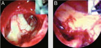
- in the treatment of neurovascular conflicts in the cerebellopontine angle with trigeminal and glossopharyngeal neuralgia (Fig. 1);
- in the treatment of extensive as well as cystic lesions in the basal and parapineal cisterns and in the third ventricle (Fig. 2).
Endoscopic control of the microsurgical process is also of great benefit in transphenoidal access to lesions in the Turkish saddle and adjacent areas (Fig. 3).
One of the most important functions of endoscopically assisted microneurosurgery (EAM) is to treat aneurysms, especially in the posterior wall of the internal carotid artery (Figs. 4 and 5) and posterior circulation (Fig. 6).
Endoscopically assisted microneurosurgery (EAM) usually uses endoscopes with a lens at an angle of 0 ° and 30 °. In the treatment of large and cystic lesions, endoscopic observation is carried out gradually by inserting the instrument "manually" during the operation. In the case of cerebral aneurysms, the endoscope should be secured with an appropriate system of holders in the operating field, and, if possible, through access that is not used in the microsurgical method. The only way to avoid damage to important vascular structures in the affected area (perforating branches) is to combine timely endoscopic and microsurgical control.
Professional training in these neurosurgeon skills is crucial for the combination of endoscopy and microsurgery. Strategic placement of monitors in the operating room is also required. The availability of specific endoscopes and holder systems is essential for achieving good results in endoscopically assisted brain microneurosurgery (EAM).
See also
- Diagnostic optics for endoscopically assisted mcroneurosurgery
- Endonasal endoscopic removal of neoplasms in the nose and paranasal cavities
- Endoscopic pituitary surgery
- Endoscopic transnasal neurosurgery
- Endoscopically assisted microneurosurgery (EAM)
- Ethmoid-pterygo-sphenoidal endoscopic approach (EPSea)
- Extended endonasal approach (EEA) in skull base surgery
- Minimally invasive microscopic surgery of the lateral skull base
- Neuroendoscope:
- Neuroendoscopy
- Neuroendoscopy in pediatrics
- Optical retractor for endoscopically assisted collection a. temporalis superficialis and cranioplasty
- Transphenoidal skull base microsurgery


