
Spontaneous intracranial (subarachnoid) and intracerebral hemorrhage
Spontaneous Intracranial Hemorrhage: Overview
Spontaneous intracranial hemorrhage (ICH) refers to bleeding within the cranial cavity that is not caused by trauma [1]. It is a major subtype of stroke, often termed hemorrhagic stroke [1]. This bleeding can occur in different locations [1, 2]:
- Intracerebral Hemorrhage (ICH): Bleeding directly into the brain parenchyma (tissue).
- Subarachnoid Hemorrhage (SAH): Bleeding into the subarachnoid space, the area between the brain and the membranes covering it, where cerebrospinal fluid (CSF) circulates.
- Intraventricular Hemorrhage (IVH): Bleeding into the brain's ventricular system (often an extension from ICH or SAH).
- Subdural or Epidural Hemorrhage: Usually traumatic, but rare spontaneous cases can occur (less common focus here).
The location, size, and underlying cause of the hemorrhage determine the clinical presentation and prognosis [1]. Common causes vary depending on the type of hemorrhage. For instance, ruptured saccular aneurysms are the most frequent cause of spontaneous SAH, while chronic hypertension is the leading cause of spontaneous deep ICH [1, 2]. Vascular malformations like arteriovenous malformations (AVMs) can cause both ICH and SAH [1].
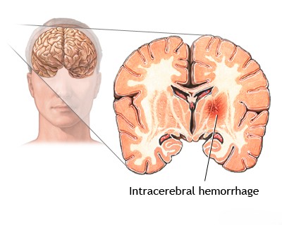

Less frequent causes include bleeding disorders, rupture of mycotic (infectious) aneurysms, hemorrhage into brain tumors, hemorrhagic transformation of an ischemic stroke, and rare vascular conditions [1, 2]. Brainstem hemorrhages can also occur due to transtentorial herniation (Duret hemorrhages) [1].
Causes of Spontaneous Intracranial Hemorrhage
The specific causes often correlate with the type and location of bleeding [1, 2]:
Primary Causes of Intracerebral Hemorrhage (ICH):
- Hypertensive Vasculopathy: Chronic hypertension damages small penetrating arteries (arteriolosclerosis, lipohyalinosis), leading to rupture, typically in deep brain structures (basal ganglia, thalamus, pons, cerebellum). This is the most common cause of spontaneous ICH.
- Cerebral Amyloid Angiopathy (CAA): Deposition of amyloid protein in the walls of small-to-medium cortical and leptomeningeal arteries, making them fragile. Typically causes lobar hemorrhages (bleeding in the lobes near the brain surface) in older individuals, often recurrent.
| Type | Most Common Cause | Typical Location | Key Clinical Clue |
|---|---|---|---|
| Intracerebral Hemorrhage (ICH) | Chronic hypertension | Deep (basal ganglia, thalamus, pons, cerebellum) | Sudden focal deficit + severe hypertension |
| Subarachnoid Hemorrhage (SAH) | Aneurysm rupture | Basal cisterns, sulci | Thunderclap headache ± meningismus |
| Lobar ICH | Cerebral amyloid angiopathy (elderly) | Cortical/subcortical lobes | Recurrent lobar bleeds + microbleeds on GRE/SWI |
| ICH/SAH from malformation | AVM or cavernoma | Variable (often lobar or deep) | Younger patient + flow voids on MRI |
Primary Cause of Spontaneous Subarachnoid Hemorrhage (SAH):
- Rupture of Intracranial Aneurysms: Weakened outpouchings on cerebral arteries rupture, releasing blood directly into the CSF-filled subarachnoid space.
- Saccular ("berry") Aneurysms: Most common type, typically at vessel bifurcations in the Circle of Willis.
- Giant Aneurysms: Large aneurysms (≥25 mm) can rupture or cause mass effect.
- Mycotic Aneurysms: Caused by infection weakening the vessel wall.
- Aneurysm rupture can sometimes lead to associated ICH or IVH in addition to SAH. Re-rupture is a major risk after initial SAH if the aneurysm isn't secured.
Causes Leading to Both ICH and/or SAH:
- Vascular Malformations:
- Arteriovenous Malformations (AVMs): Tangles of abnormal arteries and veins prone to rupture.
- Cavernous Malformations (Cavernomas): Clusters of abnormal, leaky capillaries, usually causing ICH.
- Dural Arteriovenous Fistulas (DAVFs): Abnormal connections between dural arteries and veins/sinuses.
- Hemorrhage into Brain Tumors: Both primary (e.g., glioblastoma) and metastatic tumors can bleed.
- Hemorrhagic Transformation of Ischemic Stroke: Bleeding into an area of prior ischemic stroke, especially after reperfusion therapy or with large embolic infarcts.
- Cerebral Venous Sinus Thrombosis (CVST): Blockage of venous drainage can lead to venous hypertension, causing both ischemic and hemorrhagic infarcts.
Other / Systemic Causes:
- Coagulation Disorders / Anticoagulant Therapy: Conditions like hemophilia, thrombocytopenia, liver disease, disseminated intravascular coagulation (DIC), or therapeutic anticoagulation (warfarin, DOACs) or antiplatelet use increase bleeding risk.
- Hematologic Malignancies: Leukemia, aplastic anemia can cause bleeding due to low platelets or coagulation factor abnormalities.
- Inflammatory/Infectious Conditions: Vasculitis (inflammation of blood vessels), infective endocarditis (leading to mycotic aneurysms), certain types of encephalitis (e.g., herpes simplex encephalitis, acute necrotizing hemorrhagic encephalopathy).
- Substance Abuse: Stimulant use (cocaine, amphetamines) can acutely raise blood pressure and trigger hemorrhage.
- Rare Causes: Bleeding after certain procedures (angiography, neurosurgery), associated with Moyamoya disease, radiation-induced vasculopathy, severe hypoxemia, complications of carotid-cavernous fistula (CCF), certain toxins or venoms.
Differential Diagnosis of Spontaneous Intracranial Hemorrhage [1, 2]
| Hemorrhage Type / Cause | Typical Location | Key Features / Diagnostic Clues |
|---|---|---|
| ICH - Hypertensive | Deep: Basal ganglia, thalamus, pons, cerebellum. | History of chronic hypertension. CT shows deep hematoma. |
| ICH - Cerebral Amyloid Angiopathy (CAA) | Lobar (cortical/subcortical). | Elderly patient (>60-65). Often multiple/recurrent lobar bleeds. MRI (GRE/SWI) shows lobar microbleeds, siderosis. |
| SAH - Aneurysmal Rupture | Subarachnoid space (basal cisterns, sulci). May have associated ICH/IVH. | Sudden "thunderclap" headache. CT shows SAH. CTA/DSA identifies aneurysm. |
| Vascular Malformation Rupture (AVM, Cavernoma) | ICH location depends on malformation site. AVMs can also cause SAH/IVH. | Often younger patients. MRI (AVM: flow voids; Cavernoma: "popcorn" + hemosiderin) and/or Angiography (AVM) identify lesion. |
| Hemorrhage into Tumor | Location of tumor (primary or metastasis). | MRI with contrast shows hemorrhage associated with enhancing mass. History of cancer (for mets). |
| Hemorrhagic Transformation of Ischemic Stroke | Within an area of prior ischemic infarction (vascular territory). | Occurs days after ischemic stroke onset. Imaging shows blood within established infarct. |
| Anticoagulant / Coagulopathy Related ICH | Can occur anywhere (deep or lobar). May be larger/expand. | History of anticoagulant use or bleeding disorder. Abnormal coagulation tests (INR, PTT, platelets). |
| Mycotic Aneurysm Rupture | Often distal, peripheral ICH or SAH over convexity. | Associated with infective endocarditis/sepsis. Fever common. Angiography shows distal aneurysm(s). Blood cultures positive. |
| Cerebral Venous Thrombosis (CVT) | Can cause hemorrhagic venous infarcts (often bilateral or not respecting arterial territories). | MRV/CTV confirms sinus/vein thrombosis. MRI shows infarct +/- hemorrhage. |
| Vasculitis | Rare cause. Can cause ICH or ischemic stroke. Often systemic symptoms. | Inflammatory markers. Angiography may show vessel irregularities. Biopsy needed if suspected. |
Treatment of Spontaneous Intracranial Hemorrhage
The management of ICH is an acute medical emergency. According to the latest 2025 guidelines from the American Heart Association and American Stroke Association (AHA/ASA), rapid diagnosis and intervention are critical to limiting hematoma expansion and reducing mortality and long-term morbidity [5]. Treatment pathways are broadly divided into medical management and surgical intervention.
Medical Management
Medical intervention focuses on stabilizing the patient, arresting active bleeding, and preventing secondary brain injury caused by swelling or increased intracranial pressure (ICP).
- Blood Pressure Control: Elevated blood pressure is a primary driver of ongoing bleeding. The AHA/ASA guidelines strongly recommend acute lowering of systolic blood pressure to a target of 140 mm Hg (within a range of 130–150 mm Hg) using titratable intravenous medications (e.g., nicardipine, labetalol, or clevidipine). This must be initiated as soon as possible after symptom onset [5].
- Reversal of Anticoagulation: For patients taking blood thinners, immediate reversal is paramount. Depending on the agent, this may involve administering Vitamin K, Prothrombin Complex Concentrates (PCCs), Idarucizumab (for dabigatran), or Andexanet alfa (for Factor Xa inhibitors) [5].
- ICP Management: If clinical signs of elevated ICP or brain herniation are present, acute measures such as elevating the head of the bed, administering hyperosmolar therapy (e.g., mannitol or hypertonic saline), and controlled hyperventilation are indicated [5].
- Seizure Prophylaxis: Prophylactic antiseizure medications are generally not recommended unless clinical seizures occur or the patient shows continuous epileptiform discharges on an EEG [5].
Surgical Intervention
The decision to operate on an ICH depends heavily on the hematoma's location, size, and the patient's neurological status.
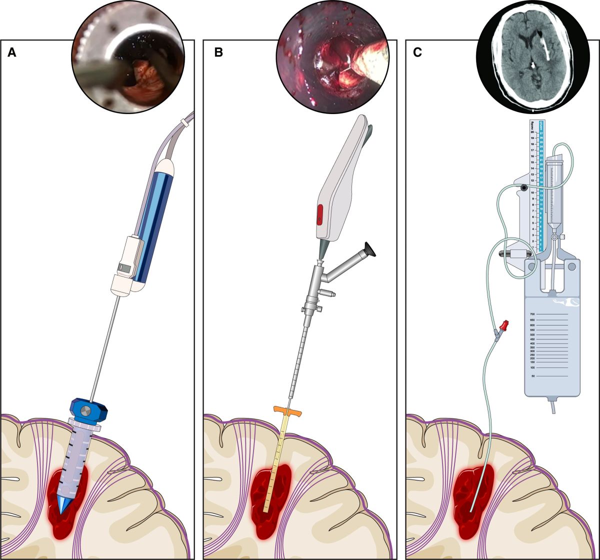
- Minimally Invasive Surgery (MIS): There is now strong evidence supporting the use of minimally invasive endoscopic evacuation for certain patients. According to the 2025 AHA/ASA guidelines, MIS evacuation of supratentorial (lobar or deep) ICH, particularly when performed within 24 hours using advanced imaging and stereotactic navigation, significantly improves functional outcomes and reduces mortality compared to medical management alone [5].
- Traditional Craniotomy: Open surgical evacuation (craniotomy) may still be indicated for large, superficial lobar hemorrhages causing massive midline shift or life-threatening brain compression [5].
- Cerebellar Hemorrhage Evacuation: A cerebellar hemorrhage measuring >3 cm in diameter, or one that is compressing the brainstem or causing obstructive hydrocephalus, requires urgent surgical evacuation to prevent fatal herniation [5].
- Ventricular Drainage (EVD): For patients with significant Intraventricular Hemorrhage (IVH) causing hydrocephalus or severely depressed consciousness, placing an External Ventricular Drain (EVD) is critical to relieve fluid pressure [5].
References
- Ropper AH, Samuels MA, Klein JP, Prasad S. Adams and Victor's Principles of Neurology. 11th ed. McGraw Hill; 2019. Chapter 34: Cerebrovascular Diseases.
- Grotta JC, Albers GW, Broderick JP, et al. Stroke: Pathophysiology, Diagnosis, and Management. 7th ed. Elsevier; 2021. Chapter on Intracerebral Hemorrhage & Chapter on Subarachnoid Hemorrhage.
- Greenberg MS. Handbook of Neurosurgery. 9th ed. Thieme; 2019. Chapter 36: Spontaneous Intracerebral Hemorrhage.
- Osborn AG, Hedlund GL, Salzman KL. Osborn's Brain: Imaging, Pathology, and Anatomy. 2nd ed. Elsevier; 2017. Section on Intracranial Hemorrhage.
- Greenberg SM, Ziai WC, Cordonnier C, et al. 2025 Guideline for the Management of Patients With Spontaneous Intracerebral Hemorrhage: A Guideline From the American Heart Association/American Stroke Association. Stroke. 2025;56(1). https://doi.org/10.1161/STR.0000000000000529
See also
- Cerebrovascular diseases - ischemic stroke, transient ischemic attack (TIA):
- Ischemic brain disease:
- Asymptomatic carotid bifurcation stenosis with noise
- Atherosclerotic thrombosis
- Atherothrombotic occlusion of basilar artery
- Atherothrombotic occlusion of internal carotid artery
- Atherothrombotic occlusion of posterior cerebral artery
- Atherothrombotic occlusion of vertebrobasilar and posterior cerebral arteries
- Atherothrombotic occlusion of vertebral and posterior inferior cerebellar arteries (PICA)
- Cerebral embolism
- Other causes of ischemic stroke (cerebral infarction)
- Small-vessel stroke (lacunar infarction)
- Ischemic stroke, transient ischemic attack (TIA), cerebral ischemia
- Sigmoid sinus suppurative thrombophlebitis with thrombosis
- Spontaneous intracranial (subarachnoid) and intracerebral hemorrhage:
- Arteriovenous malformations of the brain
- Cerebral arteries inflammatory diseases (cerebral arteritis)
- Cerebral vasospasm
- Communicating hydrocephalus after intracerebral hemorrhage with ruptured aneurysm
- Giant intracranial aneurysms
- Hypertensive intracerebral hemorrhage
- Lobar intracerebral hemorrhage
- Mycotic intracranial aneurysms
- Other causes of intracerebral hemorrhage
- Repeated cerebral artery aneurysm rupture
- Saccular aneurysm and subarachnoid hemorrhage
- Vertebrobasilar insufficiency (VBI) with vertigo symptom
Need Expert Advice on Your Condition?
Get a personalized video breakdown and treatment plan from our senior neurosurgeons using your MRI/CT scans.
👉 Request MRI Review